Sacrococcygeal Teratoma

  • Etiology: Presacral germ cell tumor consisting of totipotential cells of Hensen’s node (caudal cell mass migrates anteriorly to coccyx)
  • Imaging: mass is cystic / solid / mixed, may have increased vascularity, may have calcification
  • Types: based on location in relationship to pelvis
    — Type I – external
    — Type II – external with intrapelvic component
    — Type III – primarily internal with external component
    — Type IV – entirely internal
  • DDX: presacral mass
  • Complications: may transform into malignancy
  • Treatment: surgery
  • Clinical: AFP elevated, most common solid tumor in neonates, most common congenital neoplasm, 90% benign at birth, can present with high output congestive heart failure, may have hydrops fetalis particularly when solid, may be part of Currarino triad

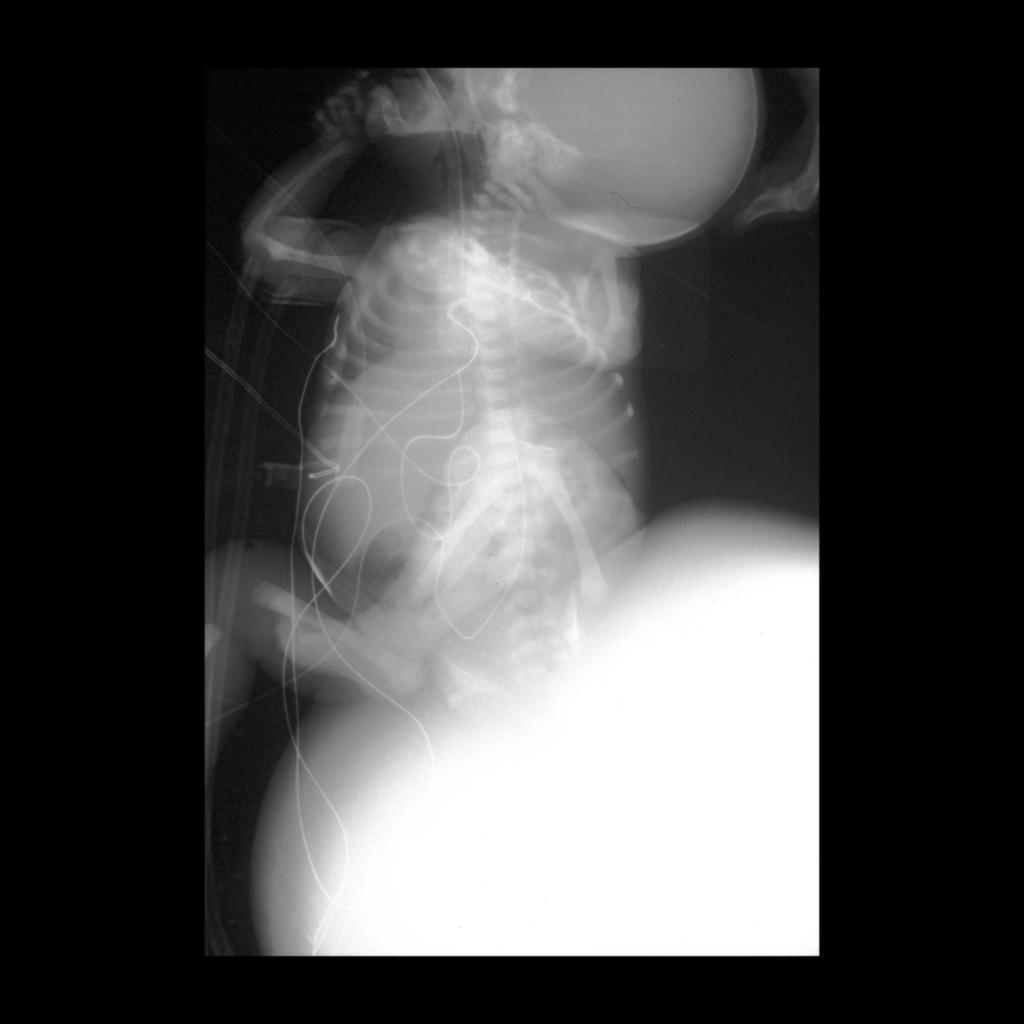
Radiology Cases of Sacrococcygeal Teratoma

Radiograph of sacrococcygeal teratoma
AXR shows a large soft tissue mass arising in the region of the sacrum.
Barium enema of presacral sacrococcygeal teratoma
Lateral image from a barium enema shows displacement of the rectum anteriorly by a presacral mass which is eroding into the rectum and causing the extravasation of a small amount of contrast from the rectum posteriorly.

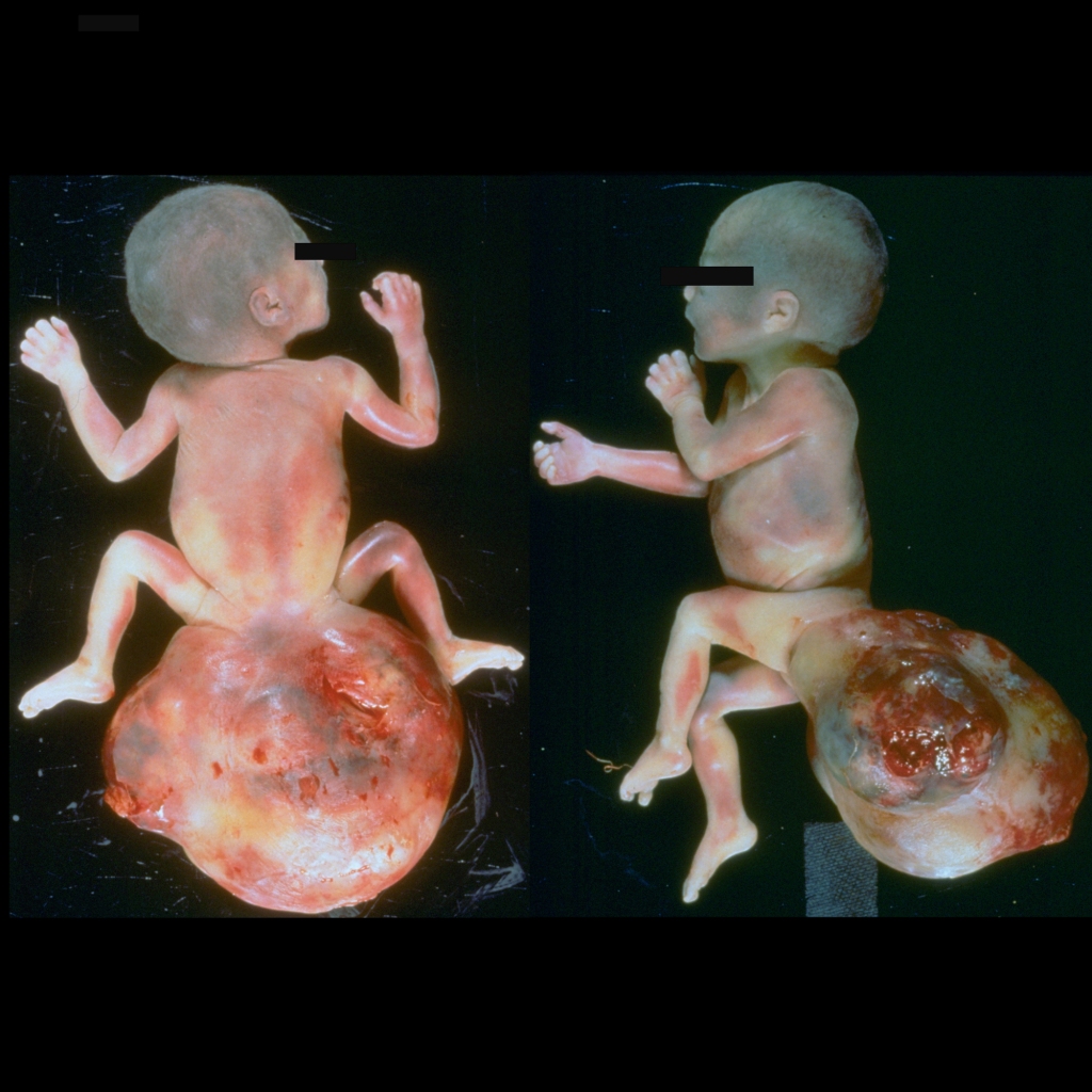
Clinical Cases of Sacrococcygeal Teratoma

Clinical image of sacrococcygeal teratoma
Clinical image shows a large exophytic soft tissue mass in the region of the sacrum.
Clinical image of sacrococcygeal teratoma
Clinical image shows a large exophytic soft tissue mass in the region of the sacrum.

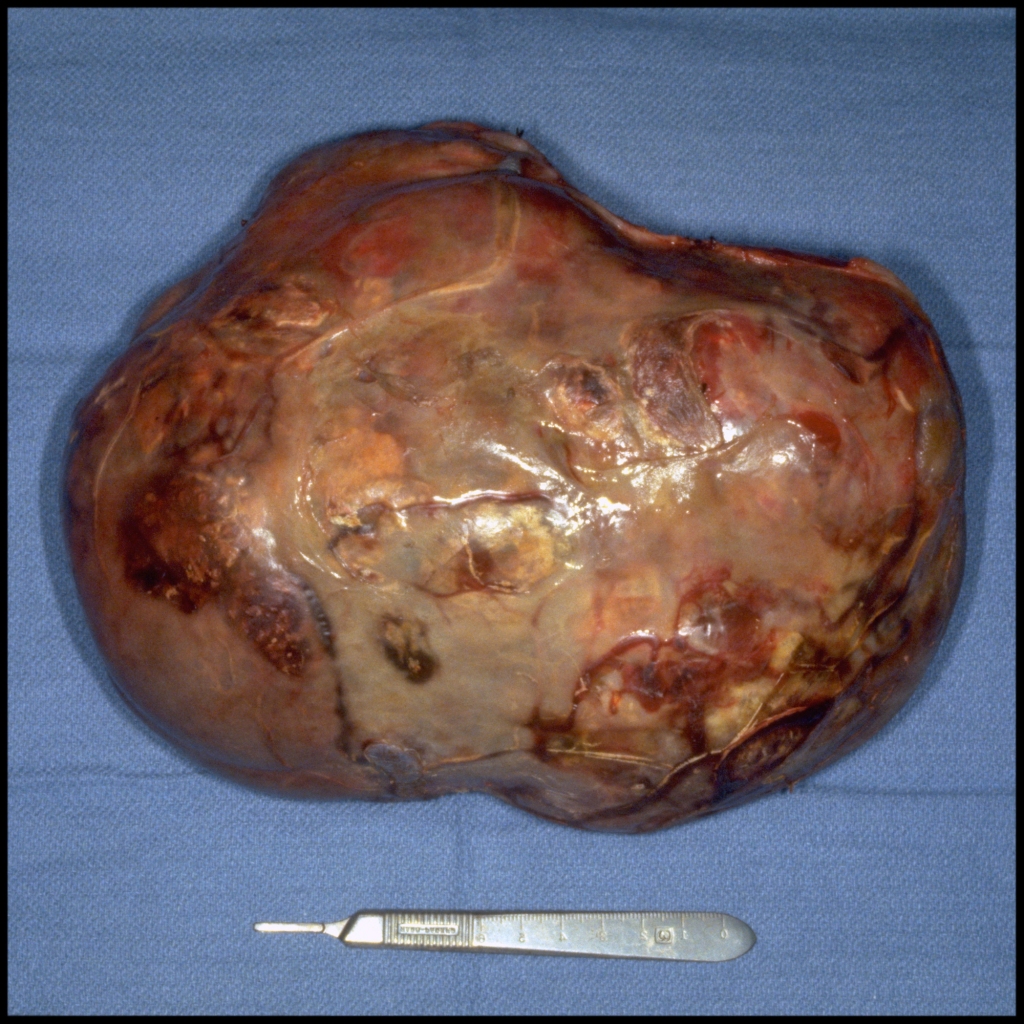
Gross Pathology Cases of Sacrococcygeal Teratoma

Gross pathology image of sacrococcygeal teratoma
Postmortem gross pathological images show a massive tumor arising from the sacrococcygeal region.
Gross pathology image of sacrococcygeal teratoma
Postmortem open (left) and sagittally sectioned (right) gross pathological images show the extent of the intrapelvic extension of the massive tumor arising from the sacrococcygeal region.
Pathological image of sacrococcygeal teratoma
Gross pathological image shows a large heterogenous sacral mass after resection.
Gross pathology image of sacrococcygeal teratoma
Gross pathological images (above) and cut gross pathological images (below) show the lesion to have a variable appearance with soft, tan areas, multiple small cysts and foci of hemorrhage. No hair or teeth were seen. A single soft tissue focus that resembled brain tissue was noted. Microscopic foci of embryonal carcinoma and endodermal sinus tumor were noted. The findings were consistent with a diagnosis of immature teratoma.

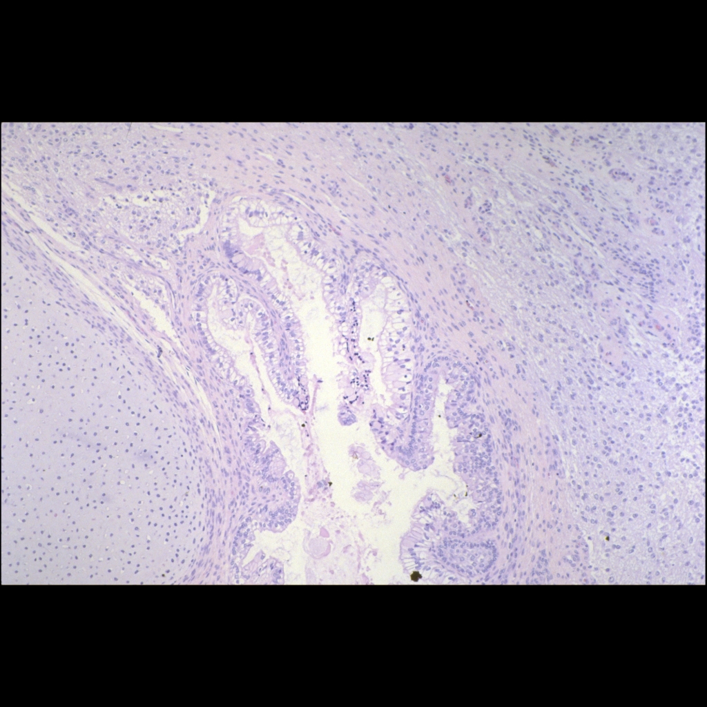
Histopathology Cases of Sacrococcygeal Teratoma

Histopathology image of sacrococcygeal teratoma
Histopathological image H&E stained slide shows the disorganized presence of all three germ cell layers – cartilage (mesoderm), space lined by ciliated respiratory type epithelium (endoderm) and neuroglial tissue (ectoderm).