Photo credit: Google
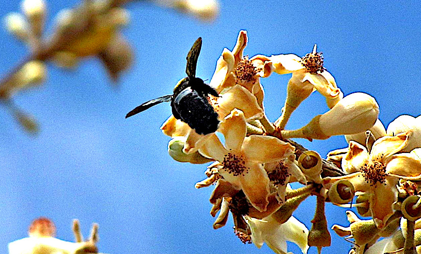
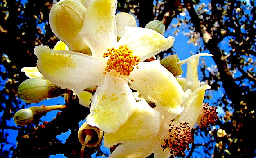
Eriotheca pubescens
Pollen and stomata morphometrics and polyploidy in Eriotheca (Malvaceae‐Bombacoideae)
by Marinho R. C., Mendes‐Rodrigues C., Bonetti A. M., Oliveira P. E., Peeters T. (2014)
Programa de Pós-graduação em Genética e Bioquímica, INGEB, Universidade Federal de Uberlândia, Uberlândia, Brazil.
===
in Plant Biology 16(2): 508 – 511 – DOI 10.1111/plb.12135 –
https://www.infona.pl/resource/bwmeta1.element.wiley-plb-v-16-i-2-plb12135
https://www.ncbi.nlm.nih.gov/pubmed/24341784
Abstract
Approximately 70% of the angiosperm species are polyploid, an important phenomenon in the evolution of those plants. But ploidy estimates have often been hindered because of the small size and large number of chromosomes in many tropical groups. Since polyploidy affects cell size, morphometric analyses of pollen grains and stomata have been used to infer ploidy level.
Polyploidy is present in many species of the Cerrado, the Neotropical savanna region in Central Brazil, and has been linked to apomixis in some taxa. Eriotheca gracilipes and Eriotheca pubescens are common tree species in this region, and present cytotypes that form reproductive mosaics. Hexaploid individuals (2n = 6x = 276) are polyembryonic and apomictic, while tetraploid and diploid individuals (2n = 2x = 92, 2n = 4x = 184) are sexual and monoembryonic.
We tested whether morphometric analysis can be used to estimate ploidy levels in E. gracilipes and E. pubescens individuals. Pollen material from diploid and hexaploid individuals of E. gracilipes, and tetraploid and hexaploid individuals of E. pubescens, were fixed in 50% FAA, and expanded leaves were dried in silica gel.
Pollen grains and stomata of at least five individuals from each population were measured. The results demonstrate that all measures were significantly different among cytotypes. Individuals with higher levels of ploidy (hexaploid) all presented measurements that were higher than those with lower levels (diploid and tetraploid).
There was no overlap between ploidy levels in each species at 95% confidence interval. Thus, the size of the pollen grains and stomata are effective parameters for analysis of ploidy levels in E. gracilipes and E. pubescens.

You must be logged in to post a comment.