Stomatal characters of the Loranthaceae in Nigeria

Fig. 1:Photomicrographs of epidermal layers of leaves of the genera Agelanthus, Englerina, Globimetula andHelixanthera in Nigeria.Key: str = striations; rh = rhaphides

Taxonomic Significance of Leaf Epidermal Characters of the Family Loranthaceae in Nigeria

by Ibrahim J. A., Ayodele A. E. (2013)

===

In World Applied Sciences Journal 24(9): 1172-1179 – DOI: 10.5829/idosi.wasj.2013.24.09.13125

https://www.academia.edu/7807047/Taxonomic_Significance_of_Leaf_Epidermal_Characters_of_the_Family_Loranthaceae_in_Nigeria?email_work_card=view-paper

 World Appl. Sci. J., 24 (9): 1172-1179, 2013
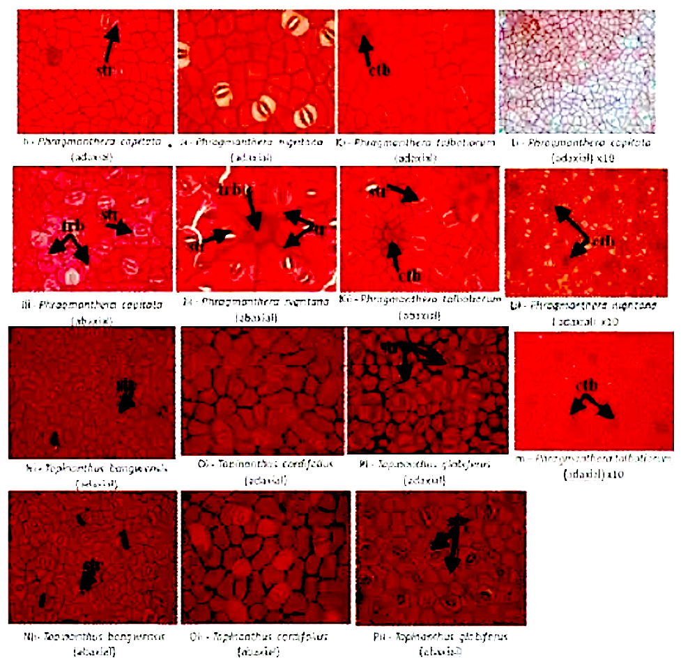
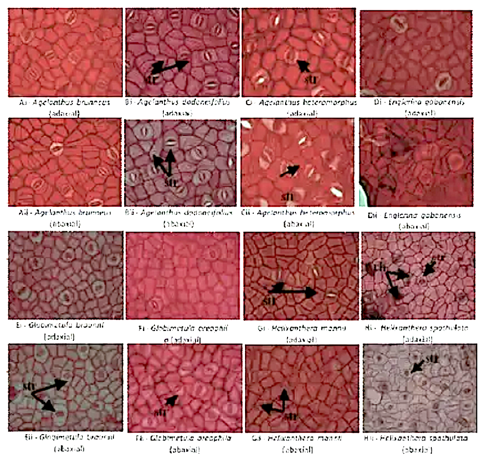
1175Fig. 1:Photomicrographs of epidermal layers of leaves of the genera Agelanthus, Englerina, Globimetula andHelixanthera in Nigeria.Key: str = striations; rh = rhaphidesFig. 2:Photomicrographs of epidermal layers of leaves of the genera Phragmanthera and Tapinanthus in Nigeria.Key: str = striations; tr = trichome; trb = trichome base; ctb = epidermal cells surrounding trichome base

Abstract

Members of the parasitic family Loranthaceae are known for their destructive nature to host plants as well as for their medicinal value. Taxonomic revision and distribution study of the family in Nigeria was recently carried out.

The taxonomic importance of leaf epidermal characters in the family Loranthaceae has been investigated with light microscopy in the present study. Amphistomatic leaf type, polygonal cell shape, straight to curved anticlinal wall and pericytic stomata types were features common to all the species.

Trichome and trichome bases were restricted to the genus Phragmanthera, striations were absent in Agelanthus brunneus (Engl.) Balle & Halle , Englerina gabonensis (Engl.) Balle and Tapinanthus cordifolius Polh. & Wiens. 

Largest cells of 67.0µm occurred in Agelanthus dodoneifolius (DC) Polh. & Wiens while the smallest cells of 27.5µm occurred inTapinanthus bangwensis (Engl. & K. Krause) Danser. 

This is the first detailed account of epidermal and stomatal characters of the Loranthaceae in Nigeria.

Stomata in Dialium (Fabaceae: Caesalpinoideae)


Epidermal morphology of the genus Dialium (Fabaceae: Caesalpinoideae)

by Sheteolu A. O., Ayodele A. E. (1997)

in Feddes Repertorium 108(3-4): 151–158 –

http://onlinelibrary.wiley.com/doi/10.1002/fedr.19971080305/full

Abstract

Epidermal morphology of adaxial and abaxial leaflet surfaces of the five species in the genus Dialium (Fabaceae: Caesalpinoideae) were studied by light microscopy.

The epidermises are composed of cells which are isodiametric, irregular or both types in outline. Anticlinal walls are either straight, curved or undulate.

Leaflets of all taxa are hypostomatic. Paracytic stomata occur in all species except D. guineense that posses both paracytic and staurocytic stomata.

Other features of the epidermis that show variation include stomatal size and density, size and density of epidermal cells, and type, size, shape, frequency and basal cells of the trichomes. An indented, dichotomous key for identifying the species is presented.

Stomata in Bignoniaceae

Plate 6: Scanning Electron Micrograph of the leaves of Bignoniaceae in Nigeria.

(A). Tecoma capensis (lower surface) showing raised stomata curved cell walls and striations on the epidermis; (B). Tecoma capensis (upper surface) showing non-glandular trichomes, with pointed ends, wavy cell walls and striations on the epidermis.


Screen Shot 2017-11-27 at 10.26.46

Plate 1: Scanning Electron Micrograph of the leaves of Bignoniaceae in Nigeria.

(A). Crescentia cujete (lower surface) showing raised stomata (Anomocytic type), peltate trichome and wavy anticlinal cell walls. (B). Crescentia cujete (upper surface) showing peltate trichome, wavy anticlinal cell walls and no stomata. (C). Jacaranda mimosifolia (lower surface) showing peltate trichome, sunken stomata and wavy anticlinal cell walls. (D). Jacaranda mimosifolia (upper surface) showing curved anticlinal walls pattern and no stomata.

Ultra-Structure of the Leaf Surfaces of the Family Bignoniaceae Juss

by Ugbabe G. E., Ayodele A. E., Kalpana S. J., Okogun J. L (2014)

G.E. Ugbabe1, A.E. Ayodele2, S.J. Kalpana3 and J.I. Okogun1

1 Medicinal Plant Research and Traditional Medicine (MPR & TM) Department National Institute for Pharmaceutical Research and Development (NIPRD), Idu-Industrial Area, P.M.B 21, Garki-Abuja, Nigeria

2 Botany Department, University of Ibadan, Ibadan, Nigeria

3 Microbiology & Biotechnology Department, Sinhgad College of Engineering (SCOE), Vadgaon (Budruk) Pune – 411 041, India

===============


in Global Journal of Botanical Science, 2014, 2, 37-44- DOI: http://dx.doi.org/10.12974/2311-858X.2014.02.01.6

GJBSV2N1A6-Ugbabe.pdf

Screen Shot 2017-11-27 at 10.29.04

Plate 2: scanning Electron Micrograph of the leaves of Bignoniaceae in Nigeria.

(A). Kigelia Africana (lower surface) showing raised diacytic stomata and curved cell walls; (B). Kigelia Africana (lower surface) showing curved anticlinal cell walls and no stomata; (C). Markhania tomentosa (lower surface) showing long non-glandular trichomes, peltate trichomes raised stomata and wavy cell walls; (D). Markhania tomentosa (upper surface) showing peltate trichome, and no stomata.


Abstract: 

The leaf surfaces of the Nigerian Bignoniaceae were studied using the Scanning Electron Microscope (SEM). The species in this study were: Crescentia cujete Linn. Jacaranda mimosifolia D. Don., Kigelia africana (Lam) Benth. Markhamia tomentosa (Benth) K. Schum., Newbuldia laevis (P. Beauv.) Seemann ex Bureau. Spathodea campanulata P. Beauv. Stereospermum acuminatissimum K. Schum. Stereospermum kunthianum Cham.Tabebuia rosea (Berthol) D. C. Tecoma stans (Linn) H, B &K. and Tecoma capensis.

The study was conducted using anatomical characters to identify and delimit the Nigerian taxa at the tribal level.

Stomata were observed on the abaxial surfaces of all the species studied. Sunken stomata were found in K. africana while the others had raised stomata.

Peltate trichomes were found on some species like the abaxial surfaces of C. cujete, J. mimosifolia, M. tomentosa, N. laevis T. stans and T. rosea and on the adaxial surfaces of C. cujete, M. tomentosa, N. laevis, S.acuminatissimum and T. roseaM. tomentosa had both glandular and non-glandular trichomes on the abaxial surface while T. capensis had only non-glandular trichome on the adaxial surface. The non-glandular trichomes of M. tomentosa were long and with blunt tip while those of T. capensis were short and with pointed tips.

Striae were found on the abaxial surfaces of J. mimosifolia and T. stans and on both the abaxial and adaxial surfaces of T. capensis.

The genera Tabebuia and Tecoma are suggested to be retained in the tribe Tecomeae based on striations on their epidermal cells. While other members be assigned to a new tribe ‘Spathodeae’.

Stomata in Andrographis (Acanthaceae)

Photo credit : Google

Andrographis paniculata

Foliar Epidermal Morphology and Anti-Diabetic Property of Andrographis paniculata (Burm. f.) Wall ex. Nees

by Fasola T. R., Ayodele A. E., Odetola A. A., Umotok N. E. (2010)

T. R. Fasolaa a , A. E. Ayodelea a, A. A. Odetolab b, and N. E. Umotoka a

a Department of Botany and Microbiology, Faculty of Science, University of Ibadan, Ibadan, Nigeria

b Department of Biochemistry, Faculty of Basic Medical Sciences, College of Medicine, University of Ibadan, Ibadan, Ibadan


in Ethnobotanical Leaflets 14: 648-53 –

http://opensiuc.lib.siu.edu/cgi/viewcontent.cgi?article=1674&context=ebl


Abstract

The leaf epidermal features and anti diabetic property of Androgragphis paniculata were investigated.

The epidermal cells are polygonal to irregular with curved to undulate anticlinal walls on both surfaces of the leaf.

The leaf is hypostomatic with stomata restricted to the abaxial surface. Stomata type is axillocytic.

The epidermal cells contain deposits of crystal oxalates while cystoliths are found in a few cells. The leaf extract was evaluated for hypoglycemic effects. There was significant hypoglycemic effect in alloxan induced diabetic rats following administration of the leaf aqueous extract. The result shows that the level of fasting blood sugar was dependent on the previous level of basal blood sugar in the four groups of rats used for this study. Treatment of the rats with A. paniculata showed a dose dependent relationship with the time it took for the blood of the rats to return to the basal blood sugar levels. The study supports the use of the plant in the management and treatment of diabetes.

Stomata in Uapaca (Euphorbiaceae)

Photo credit: Google

Uapaca togoensis

Comparative leaf epidermal morphology of five West African species of Uapaca baill (Phyllanthaceae pro forma Euphorbiaceae)

by Kadiri A. B., Ayodele A. E., Olowokudejo J. D., Uchemefuna D. (2013)

in Nigerian Journal of Botany 2013

– http://nijbot.org/?m=201309&paged=2

Foliar epidermal features of the species of Uapaca. A, B: U. heudelotii; C, D: U. paludosa; E, F: U. staudtii. Note the straight anticlinal wall and the polygonal cells. A,C, E: Adaxial surface and B, D, F: Abaxial surface. Bar scale is 50µm. - http://nijbot.org/wp-content/uploads/2013/09/Images.jpg
Foliar epidermal features of the species of Uapaca. A, B: U. heudelotii; C, D: U. paludosa; E, F: U. staudtii. Note the straight anticlinal wall and the polygonal cells. A,C, E: Adaxial surface and B, D, F: Abaxial surface. Bar scale is 50µm. – http://nijbot.org/wp-content/uploads/2013/09/Images.jpg

ABSTRACT
The taxonomy of Euphorbiaceae was recently reviewed based on morphology and molecular data and more data are still being produced to enhance comprehension of the component taxa.

Therefore, in this study, we have contributed additional taxonomically useful data on the leaf epidermis of Uapaca which is now in the new family segregate, Phyllanthaceae of Euphorbiaceae by the means of light microscopy.

Generic constant features are polygonal cell shape, straight anticlinal wall pattern, hypostomatic leaf, paracytic and anomocytic stomata types.

Distinguishing characteristics are absence of anisocytic stomata in U.  staudtii, presence of pericytic stomata in U. heudelotii and U. paludosa and presence of tetracytic stomata in U. vanhouttei.

Other taxonomically useful features are possession of conical non-glandular trichomes in U. togoensis on both surfaces but which are restricted to the abaxial surface in U. vanhouttei as well as presence of crystals of calcium oxalate in U. staudtii and U. togoensis.

In the genus, the stomatal index ranges from 22.4% to 34%.

The taxonomic relationship is discussed and an artificial indented dichotomous key for separating the species is presented.

Read the full article: NJB