
Biomineralization in Polychaete Annelids: A Review


Institute of Ecology and Earth Sciences, University of Tartu, Ravila 14A, 50411 Tartu, Estonia
Minerals 2021, 11(10), 1151; https://doi.org/10.3390/min11101151
Submission received: 18 August 2021
/
Revised: 14 October 2021
/
Accepted: 18 October 2021
/
Published: 19 October 2021
(This article belongs to the Special Issue Biomineralization and Biominerals: Lessons from Mineral-Producing Organisms)
Abstract
:Polychaete annelids are a very important group of calcifiers in the modern oceans. They can produce calcite, aragonite, and amorphous phosphates. Serpulids possess very diverse tube ultra-structures, several unique to them. Serpulid tubes are composed of aragonite or calcite or a mixture of both polymorphs. The serpulid tubes with complex oriented microstructures, such as lamello fibrillar, are exclusively calcitic, whereas tubes with prismatic structures can be composed either of calcite or aragonite. In serpulids, the calcareous opercula also have complex microstructures. Evolutionarily, calcitic serpulid taxa belong to one clade and the aragonitic taxa belong to another clade. Modern ocean acidification affects serpulid biomineralization. Serpulids are capable of biomineralization in extreme environments, such as the deepest part (hadal zone) of the ocean. The tubes of calcareous sabellids are aragonitic and have two layers, the inner irregular spherulitic prismatic layer and the outer spherulitic layer. The tube wall of cirratulids is composed of aragonitic lamellae with a spherulitic prismatic structure. In some other polychaetes, biominerals are formed in different parts of the animal body, such as chaetae or body shields, or occur within the body as granule-shaped or rod-shaped inclusions.
1. Introduction
Calcifying polychaete annelids are a very important group of animals in the oceanic calcium sink, especially in temperate seas where they can be major calcifying invertebrates [1,2,3]. Some serpulids are economically important due to intense biofouling of artificial substrates in the sea. Isotopic analyses have been used to study the stable-isotope composition of serpulid tubes for insights into calcification processes in marine organisms [4]. Serpulid tubes have been studied with plasma mass spectroscopy (IPC-MS) to analyze the anthropogenically mobilized metal content (e.g., Al, Cd, Ni, Pb, U, etc.) as an estimate of the human impact on the coastal environment of California [5].
General understanding of the evolution of invertebrate biomineralization systems is mostly based on studies of molluscs (Carter et al. and references therein) [6], and to a lesser extent on corals, brachiopods, bryozoans, and echinoderms. Polychaetes, however, may have a different biomineralization strategy, the understanding of which would contribute significantly toward a general synthesis of biomineralogical evolution among invertebrates having calcareous skeletons.
Global warming is not the only outcome of rising levels of carbon dioxide in the atmosphere. The oceans are becoming more acidic due to anthropogenic CO2 emissions. Experiments suggest that invertebrates constructing their shells of aragonite and high Mg calcite, such as many serpulids [3], will be particularly affected by oceanic acidification.
The first analyses of polychaete biomineralization described serpulid (Figure 1) tube formation [7,8,9,10,11]. During the last decades, our knowledge of biomineralization has grown not only for serpulids [12,13,14,15,16,17,18,19,20,21,22,23,24,25,26,27,28,29,30,31] but also other polychaete annelids [32,33,34]. However, our understanding of polychaete biomineralization is far from complete and many aspects are controversial. This review summarizes the most important aspects of the polychaete biomineralization currently known.
2. Materials and Methods
All photos of serpulid tubes and tube sections, opercula, sabellid, and cirratulid tubes were taken with a scanning electron microscope (SEM). Photographed samples were polished and etched in a 1% solution of acetic acid for 1 min. All preparations were coated with gold prior to the SEM study. A Hitachi S-4300 SEM was used for taking the photos. Microscopy (SEM) was performed at the Swedish Museum of Natural History, Stockholm. The beam was operated at 5–10 kV and 1 nA during the photo session. The Hitachi S-4300 SEM, equipped with an Inca Energy Dispersive X-Ray Analysis (EDX) system, was also used to study the elemental composition of biominerals, especially contents of Sr and Mg that are indicative for aragonite and calcite. The overall mineral composition (aragonite/calcite) of polychaete tubes was studied by means of X-ray diffraction (XRD) in powdered samples on a Dron-3M diffractometer, at the Department of Geology, University of Tartu. Spectroscopic data to distinguish aragonite from calcite in cirratulid and deep-water serpulid tubes were obtained using a T64000 triple-stage laser Raman system (JY Horiba, Edison, NJ, USA) possessing macro-Raman and confocal micro-Raman capabilities.
3. Biomineralization of Serpulids
In their studies, Hedley and Neff [7,8,9,10,11] described serpulid tube formation for the first time. Neff [7] used a transmission electron microscope (TEM) to study the secretion of calcium carbonate in Spirobranchus americanus (as Pomatoceros caeruleus). He found that S. americanus produced calcareous granules of cubic or rhombohedral shape with average dimensions of 0.15–0.2 µm in its calcium-secreting glands. These secretory granules contain a fibrous organic matrix with needle-like low-magnesium calcite crystals [7]. According to Neff’s model, the worm uses the calcareous granules to build its tube. The granules are secreted as a carbonate slurry and the worm shapes and plasters it onto the tube aperture using the undersurface of the collar. Thereafter, the slurry solidifies to form a new mineral lamella of the tube. Weedon [16] suggested that some oriented tube structures are likely the result of controlled molding of the calcite-saturated mucus in forward and backward applications by the serpulid’s collar. Based on the complex oriented biomineral structures of S. americanus and many other serpulids, Vinn et al. [21,35] suggested a matrix-controlled crystallization model for serpulid tube formation analogous to that of the biomineralization of mollusc shells. The calcareous serpulid opercula are secreted by the epithelium via an organic matrix-controlled crystallization model [26,36,37,38,39,40].
3.1. Skeletal Structures
The serpulid tubes are composed of convex forward lamellae that have a chevron shape in the longitudinal section. These chevron-shaped lamellae are laid down successively from the apertural end of the serpulid tube. Both ends of chevron-shaped lamellae are sub-parallel to the surface of the tube near the outer and inner surfaces [16]. There can be two or more microstructural zones or layers parallel to the tube surface within a single chevron-shaped lamella of the serpulid tube. The majority of modern serpulids have single layered tubes, but many serpulids have two- or multi-layered tubes, and these microstructural layers cross the chevron-shaped growth lamellae. In two-layered tubes, chevron-shaped growth lamellae have a different microstructure in the external and internal parts of the lamella. Multilayered tubes are rare as compared to single- and two-layered tubes. Two-layered and multilayered serpulid tubes tend to have an outer layer composed of dense mineral structures, such as many oriented prismatic structures [41]. Serpulids possess several unique tube microstructures [42,43,44]. Some serpulids have biomineralized opercula. These opercula can contain up to two layers with distinct microstructures [26].
Fifteen distinct types of mineral microstructures have been described in serpulids [21,26,29,42]. The category of isotropic structures where the elongation axis of the crystals lacks uniform orientation comprises: (1) the irregularly oriented prismatic (IOP) structure (Figure 2), (2) the spherulitic irregularly oriented prismatic (SIOP) structure, (3) the irregularly oriented platy (IOPL) structure, (4) the homogeneous angular crystal (HAC) structure, and (5) the rounded homogeneous crystal (RHC) structure.
The category of semi-oriented structures where the elongation axis of crystals has a semi-uniform orientation comprises: (1) the semi-ordered irregularly oriented prismatic (SOIOP) structure (Figure 3), (2) the preferentially orientated prismatic (SPOP) structure, and (3) the semi-ordered spherulitic irregularly oriented prismatic (SOSIOP) structure.
The category of oriented prismatic structures where the elongation axis of the crystals has a uniform orientation and is continuous through successive growth increments comprises: (1) spherulitic prismatic (SPHP) structure (Figure 4), (2) the regular spherulitic prismatic (RSPHP) structure, (3) simple prismatic (SP) structure, and (4) the regularly ridged prismatic structure (RRP).
The category of oriented complex structures where the axis of the crystallites has a uniform orientation that is parallel to the tube wall comprises the following structures: (1) the lamello-fibrillar (LF) structure (Figure 5), (2) the spherulitic lamello-fibrillar (SLF) structure, and (3) the ordered fibrillar (OF) structure.
3.2. Organic Sheets in Mineral Structures and the Inner Organic Tube Layer
There are organic sheets (Figure 5a) in the mineral tube structure in certain serpulid species that belong to one monophyletic clade (A) that is supported by both morphological and molecular data (i.e., Crucigera, Hydroides, Serpula, Ditrupa, Pseudochitinopoma, Ficopomatus, Galeolaria, Spirobranchus, and Laminatubus), but some species in that clade do not have them. The organic sheets can likely be used in distinguishing the two major clades of serpulids. Organic sheets are very numerous and best-developed in the genus Spirobranchus. Vinn [27] suggested that organic sheets have evolved to strengthen the mechanical properties of the tubes in clade A, which also contains serpulids with complex mineral tube microstructures. Presumably, the lower surface of the organic sheet could terminate the crystal growth, whereas the upper surface is likely used for crystal nucleation. Vinn [27] suggested that the chemical properties of the upper and lower surface of the organic sheets could be different. The upper surface of organic sheets could support nucleation of crystals, whereas the lower surface of organic sheets could terminate the growth of crystals. In the case of oriented prismatic structures, the orientation of crystals on the organic sheets is controlled by heteroepitaxy (i.e., crystals have the same orientation on both sides of organic sheets), whereas in complex oriented structures where prismatic elements of the structure are parallel to the sheets, the orientation of crystals is not controlled by heteroepitaxy (by the crystal orientations of the previous growth increment). Tanur et al. [25] studied organic sheets in H. dianthus and found that they could be composed of polysaccharides.
All serpulids possess an internal organic tube lining (Figure 6) [14,21,25]. Tanur et al. [25] identified the composition of inner organic linings and found that in Hydroides dianthus, it was composed of collagen-containing fibers. The orientation of spherulitic prisms found in the innermost layer of the tube wall of recent Crucigera websteri, C. zygophora, Floriprotis sabiuraensis, and Pyrgopolon ctenactis revealed that the structure grew toward the tube’s lumen and thus towards the organic inner tube lining [45]. Analogous directions of growth for this structure were described in Hydroides sp. from the Miocene of Austria. If the organic tube lining was used for nucleation, the spherulites should grow away from the lumen [45].
3.3. Minearal Composition
Serpulid tube mineralogy does not seem to differ between marine, brackish, and fresh water environments [21]. Skeletal carbonate mineralogy has been studied only in as few as 15% of extant species (n = 52), and about half of recent genera (n = 25). Serpulid tubes vary in their skeletal mineralogy. The mineral composition of serpulid tubes has been reported to be either aragonitic, calcitic, or a combination of both [3,13,21,46,47,48]. Serpulid tubes can be entirely aragonitic (about 24% of species) to entirely high-Mg calcite (40% of species), to mixtures of the two polymorphs [3]. Mg content in serpulid calcite ranges from 7 to 15 wt.% MgCO3, with a mean of 11 wt.% MgCO3 [3]. There is little mineralogical variation within aragonitic species. On the other hand, high-Mg calcitic species have somewhat higher variability in both Mg and calcite content, and species with the mixed mineralogies are most variable [3]. The serpulids in one clade are calcitic, whereas in the other clade they are aragonitic. Thus, there is a phylogenetic control on the mineralogical composition of serpulid tubes [3].
3.4. Development and Serpulid Biomineralization
Very little is known about the ontogenetic changes in the biomineralization in serpulids. Chan et al. [28] studied the mineral composition and ultrastructure of the calcareous tubes built in various developmental stages of Hydroides elegans. They examined various early calcifying juvenile stages and the adult worm tubes using XRD, FTIR, ICP-OES, SEM, and Raman spectroscopy. They discovered ontogenetic changes in carbonate mineralogy. The juvenile tubes of H. elegans were predominantly aragonitic and contained more amorphous calcium carbonate, whereas adult tubes were always bimineralic with much more calcite [28]. They observed that the change in mineral composition was gradual. During the ontogeny of the tube, the Sr/Ca ratio decreased, and the Mg/Ca ratio increased [28]. The inner tube layer in H. elegans has a calcitic composition, whereas the outer layer is aragonitic. Similarly, the microstructural complexity of the tube increased with the tubeworms’ age. During ontogeny, the appearance of unoriented ultra-structures was followed by oriented ultra-structures, and this may repeat the order of evolution of serpulid ultra-structures from unoriented to oriented structures [28].
3.5. Serpulid Biomineralization and Environment
The calcium carbonate dissolves better in more acidic water [49]. The dissolution of CaCO3 increases with the low water temperatures, but less for calcite than for aragonite [50]. This is because cold water contains more dissolved CO2 and is more acidic than warm water. Water temperature has played an important role in the mineralogical evolution of some invertebrates, such as bryozoans [51], where cold temperatures favor calcitic composition of the skeleton or calcitic external layers. In contrast, the possible effects of water temperature on serpulid biomineralization are largely unknown. The pressure increases the solubility of CO2 in sea water and thus increases the dissolution of calcite and aragonite, with a stronger effect on aragonite [44]. The cold and corrosive waters of the deep ocean (hadal zone) have been observed to partially dissolve the serpulid tubes [44]. The calcium carbonate (aragonite and calcite) supply equals the rate of dissolution in the ocean at 4000–5000 m, which is termed the “Carbonate Compensation Depth” (CCD) [52]. The CCD is often argued to be a physiological barrier to deep ocean biomineralization for organisms with calcareous exoskeletons. However, serpulid species belonging to the genera Bathyditrupa, Bathyvermilia, Hyalopomatus, Pileolaria, and Protis have been reported from 5020 to 9735 m [44]. The tube microstructure of hadal specimens of Protis resembles that of shallow water ones—they all have an irregularly oriented prismatic tube microstructure [44]. Kupriyanova et al. [44] also did not find any specific tube microstructures in other hadal serpulids. Moreover, they discovered that, in addition to calcitic tubes (Bathyditrupa challengeri), hadal serpulids can also have aragonitic tubes (Bathyditrupa hovei and Protis sp.) [44]. The surprising discovery of aragonite in deep-sea serpulids indicates that these tubeworms are able to maintain and secrete this unstable biomineral in the deepest parts of the ocean. Kupriyanova et al. [44] suggested that a comparative investigation of the chemical composition of serpulid tubes will reveal important differences in the organic tube matrix of deep-sea and shallow-water serpulid tubes.
As a result of anthropogenic CO2-driven ocean acidification (OA), shallow seas are becoming increasingly challenging for calcifiers such as serpulid tubeworms due to reductions in saturation states of calcium carbonate (CaCO3) minerals [53]. The influence of OA on the calcification rate has been the most frequently investigated process, nevertheless, OA may affect the quality of calcareous worm tubes through weakened calcification processes even if there are no observable changes in the calcification rate [53]. Juvenile tubes of Hydroides elegans are primarily composed of the highly soluble CaCO3 polymorph, aragonite [53]. Tubes of H. elegans built in seawater with aragonite saturation states near or below one contain much higher proportions of amorphous calcium carbonate (ACC). Moreover, their calcite/aragonite ratio is markedly increased. These OA-caused alterations in the mineralogical composition of serpulid tubes result in a great decrease in the tube elasticity and hardness [53]. Therefore, the aragonite-producing juvenile H. elegans can no longer maintain the integrity of their calcification products in conditions where ΩA is near or below one. This will likely result in reduced survivorship of H. elegans due to the weakened tube protection [53,54]. In an experimental work, Chan et al. [55] found that decreased pH and reduced salinity at elevated temperature forced H. elegans to construct tubes with a more compact ultrastructure, resulting in enhanced hardness and elasticity as compared to tubes produced during decreased pH at ambient temperature. They discovered that elevated temperature neutralized the decreased pH-caused tube damage in H. elegans [56]. As for climate change, one would expect to see that serpulid tubeworms will be resilient to the projected near-future decreased pH or salinity when it is accompanied with surface seawater temperature rises of at least 4 °C [55]. Recently, Ni et al. [57] found a resistance of Spirorbis spirorbis tube growth to OA levels predicted for the year 2100 in the Baltic Sea. Their results also revealed that Baltic S. spirorbis prefer moderately warmer conditions during their early life stages, but could suffer from increasing shell corrosion as a consequence of progressing OA.
4. Biomineralization of Sabellids
Only a single species of modern sabellids, Glomerula piloseta, dwells in calcareous tubes, though there are several fossil calcareous sabellids known from the Permian to Paleogene [58,59,60]. Sabellids are phylogenetically closely related to serpulids [61]. The biomineralization process of sabellid tubes is likely similar to serpulid opercula and probably also to serpulid tubes, and resembles biomineralization in molluscs and many other invertebrate phyla. Most likely, the sabellid skeleton is formed by extracellular mineralization, mediated and controlled by an organic matrix. This organic matrix is secreted together with calcium ions by a secretory epithelium [32].
4.1. Skeletal Structures
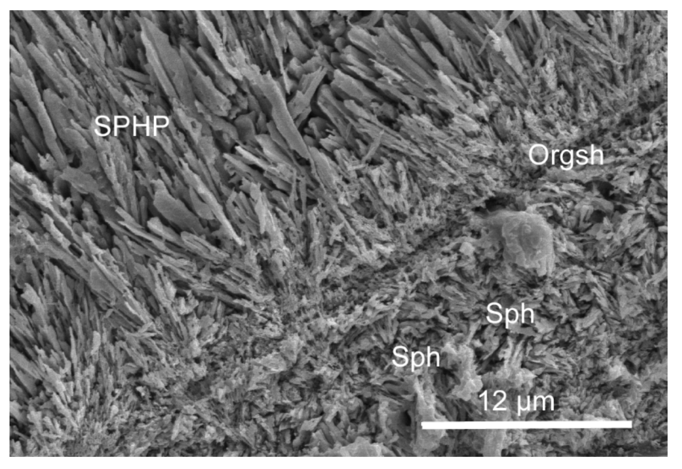
The tube structure of calcareous sabellids is simple and primitive. Their tube wall is composed of an outer layer of spherulites and an inner layer of spherulitic prisms (Figure 7) [32]. The friable outer layer of recent Glomerula piloseta is porous, c. 25 µm thick, and composed of regularly shaped small spherulites. The inner layer of the G. piloseta tube is 50–60 µm thick. The outer and inner layers are separated by thin organic films. At the contact of the two layers, bundles of needle-shaped crystallites forming an irregular spherulitic prismatic structure grow towards the tube lumen. The individual prisms in Glomerula are not encased by an organic film. The distal ends of prisms form irregular elevations on the tube lumen. The spherulitic prisms can epitaxially grow through several organic films that mark the boundaries of growth increments [32].
4.2. Mineral Composition
5. Biomineralization of Cirratulids
Only a few species of cirratulids build calcareous tubes. Fischer et al. [62] described cirratulid tubes as formed by micritic peloidal lamellae. These lamellae form a stromatolitic-like fabric that is intercalated with lenses of fibrous aragonite/calcite [62]. In their review of cirratulid biomineralization, Fischer et al. [62] concluded that these worms precipitate their hard parts by a hitherto unknown method for any eumetazoan. They described two mechanisms: (1) outside the soft tissue within a Ca2+ binding mucus, excreted by a basal layer, from which calcified lamellae are produced, and (2) calcification of bacterial rods and the remains of biofilms. This process is not controlled by the animal itself. The latter process produces peloidal aggregates and fibrous cement rims [62]. On the basis of similarities in the tube microstructure with calcareous sabellids, Vinn [33] hypothesized that tube formations of sabellids and cirratulids could be similar. The spherulitic prismatic microstructures of sabellids and cirratulids resemble some structures of molluscs. If the biomineralization of sabellids [32] and cirratulids is similar to the extracellular biomineralizers such as molluscs, for example, then their skeletons could be formed by extracellular mineralization [33]. In molluscs, extracellular mineralization is mediated and controlled by an organic matrix that is secreted together with calcium ions by a secretory epithelium [63].
5.1. Skeletal Structures
The tube wall of Dodecaceria coralii and D. caulleryi is composed of numerous thin calcareous lamellae. These lamellae are 1.5–3.0 µm thick and have a spherulitic prismatic structure (Figure 8) [33]. In some parts of the tube wall of D. caulleryi, the organic sheets between the calcareous lamellae are more heavily calcified than in the interior of the calcareous lamellae. In cirratulids, the calcareous spherulitic prisms are not epitaxially continuous through several growth lamellae, and that is different from sabellids. In Diplochaetetes mexicanus, the tube wall is also composed of lamellae with mostly spherulitic prismatic structure, but in addition, there are also lamellae with a homogeneous structure formed by unoriented calcareous rods [33].
5.2. Mineral Composition
Taylor et al. [34] used the Raman spectroscopy to study the mineralogical composition of the tubes of three calcareous cirratulid species. They found that Raman spectra of Dodecaceria caulleryi, D. coralii, and fossil Diplochaetetes mexicanus tubes are similar, indicative of pure aragonite from the presence of a low-frequency Raman band at ~205 cm−1. Point spectra of these three cirratulids and mapping of an area from D. coralii failed to identify calcite (i.e., reveal the existence of ~283 cm−1 bands), indicating that the cirratulid tubes are composed of pure aragonite. In all the EDX analyses performed by Taylor et al. [34], no or very little Mg was detected, but abundant Sr was present, corroborating the Raman spectroscopy results that indicated a pure aragonitic composition.
6. Biomineralization in Non-Tubicolous Polychaetes
In some polychaetes, biominerals are produced as a part of the animal body, such as chaetae or body shields. They are often included in the animal body as granule-shaped or rod-shaped inclusions [64]. In the opheliid species Thoracophelia minuta, approximately 300 rod-like inclusions have been found in the coelomic cavity filled with body fluid. The rod-like inclusions were black, approximately 20 μm long, had a corrugated rod shape, and some of them were curved. In a SEM–EDS analysis carried out by Jimi et al. [64], high levels of carbon, oxygen, sulfur, and calcium were detected from spicules, in contrast to chaetae, which had a high content of carbon, oxygen, and chloride.
Amphinomid fireworms are well-known for their stinging notochaetae (dorsal bristles) [65], which are calcareous. Righi et al. [65] examined the mineralogical and chemical composition, the external structure, and the ultrastructure of the ventral and dorsal chaetae in Hermodice carunculata. Their analyses clearly identified crystalline calcium carbonate and clusters of amorphous calcium phosphate in the fireworms’ stinging bristles. Tilic et al. [66] described granules with a dense core in the ultrastructure of the chaetae of E. complanata. According to their interpretation, these granules were “carbonated apatite nodules”, indicated by their pseudo-hexagonal morphology. In a XRPD and TGA study, Righi et al. [65] confirmed that CaCO3 is abundant in H. carunculata, but they found no evidence for crystalline apatite. Using energy dispersive X-ray measurements and scanning electron micrographs, Righi et al. [65] demonstrated that the dorsal chaetae have an extremely shallow insertion point in the body, in contrast to the ventral chaetae. This could facilitate the release of the notochaetae in the environment. The proximal part of each notochaeta contains canals with a hexagonal pattern rich in Ca and P [65].
7. Discussion
7.1. Serpulid Biomineralization and the Controversy
There is not much debate about the chemical composition of serpulid tubes. It has been established that serpulid tubes contain both soluble (in EDTA) and insoluble organic matrices [25], which are characteristic of biomineralization controlled by the organic matrix. The carboxylated and sulfated polysaccharides are the major components of the soluble organic matrix in serpulid tubes [25]. In addition to major components, there are various amino acids, such as aspartic acid, glutamic acid, glycine, and proline, within the soluble organic matrix in serpulid tubes. Sulfated and carboxylated polysaccharides partially control the process of biomineralization. They are capable of binding cations, and that is important for the nucleation process [63]. However, organic sheets in serpulid tubes are not always directly involved as a template in formation of complex oriented tube structures. In several species such as Serpula israelitica, S. vermicularis, and Spirobranchus triqueter, long segments (several growth increments with different crystal orientations) of LF structure could occur without any organic sheet. The occurrence of organic sheets in serpulids is similar to that of molluscs and could indicate a mollusc-type matrix-mediated biomineralization [6].
There is controversy involved in the models of the formation of serpulid tube microstructures. The classical understanding of serpulid biomineralization involves the so-called carbonate slurry model [7,8,9,10,11]. According to this model, calcareous granules, produced by the calcium-secreting glands in Spirobranchus americanus, are used to build the tube in which the animal lives. The granules are secreted as a carbonate slurry that is shaped and plastered to the tube aperture by the undersurface of the collar, and such a process must result in the formation of an unoriented microstructure. Thereafter, the slurry solidifies to form a new mineral lamella of the tube. However, serpulids possess diverse fabrics of simple oriented and complex oriented microstructures that cannot be explained by the carbonate slurry model. Weedon [16] suggested that some oriented tube structures, such as the LF structure in Spirobranchus triqueter, could result from controlled molding of the calcite-saturated mucus in forward and backward applications by the serpulid’s collar. Similar ideas were recently put forward by Buckman and Harries [30], who argued that “the difference in orientation observed between unordered and ordered fabrics can be explained by the application (or lack of) of rotational force. Ordered fabric on the inside of the tube could be produced by a combination of rotational movement of the serpulid’s ventral-shield (or possibly the operculum)/collar, combined with downward force achieved by partial retraction, allowing the alignment of calcite laths within an organic mucus”. This would offer an alternative explanation for LF-type complex oriented structures if all crystallites in the single growth increment would have the same orientation. The actual situation in many multilayered serpulids is that the same growth increment contains several zones with different microstructures (for example, SPHP, IOP, LF, and SPHP in Cruzigera websteri) and crystallites with different orientation [21]. It means that the same growth increment goes across different microstructures and it is impossible that with the same rotational movement of the serpulid collar the animal would create multiple types of microstructures and the multitude of crystallite orientations (Figure 9). Thus, one must conclude that the carbonate slurry model, even backed up with possible specific rotational activities of the serpulid collar, cannot explain the formation of LF-type structures, and the matrix-controlled crystallization remains the most plausible hypothesis for serpulid biomineralization. However, the present data are not sufficient to infer the mineralization process (i.e., induced, controlled) with certainty.
7.2. Evolution of Polychaete Biomineralization
Serpulids have the highest diversity of skeletal microstructures among polychaetes. They are the only polychaetes with complex skeletal microstructures, and therefore it is likely that their biomineralization system is more advanced than in the other polychaete annelids [21,25]. Serpulids can build tubes of calcite, aragonite, or a mixture of both of these polymorphs, whereas sabellids and cirratulids are capable of producing only aragonitic tubes [32,34]. Serpulid skeletal microstructures are similar to those found in a variety of invertebrate phyla, and the diversity and complexity of their structures is comparable to that of brachiopods and bryozoans. On the other hand, only two types of tube microstructures have been found in sabellids and cirratulids [32,33]. Serpulids form two major clades [67], where oriented microstructures, either simple or complex, occur only in one clade, whereas unoriented microstructures are found in both clades (Figure 10). The most complex serpulid tube microstructures are calcitic, and that is different from molluscs, where aragonitic structures are more complex than calcitic structures [6]. The serpulids with complex microstructures likely appeared later in the evolution than the clade with isotropic tube microstructures. The mineralogical evolution of polychaete annelids has not been investigated in detail. The earliest serpulids and sabellids presumably began to calcify in the Aragonite II seas of the Permian Period [58,68], and aragonite is presumed to be the primitive biomineral for both serpulids and sabellids [32,69]. It is possible that the dominant mineralogy of serpulids changed to calcitic during the extreme Calcite II seas of the Late Cretaceous [70]. It is interesting that the bryozoans, for example, are primitively calcitic, and they appeared during an epoch of calcitic seas (Late Cambrian), whereas both molluscs and serpulids are primitively aragonitic and appeared during an epoch of aragonitic seas in the Early Cambrian and Permian, respectively. The other groups of polychaetes played only minor roles in the evolution of polychaete biomineralization, and their biomineralization systems are primitive as compared to serpulids.
8. Conclusions
Fifteen tube microstructures occur in serpulids, whereas in sabellids and cirratulids, only two types of tube microstructures have been described. Serpulids have the most advanced biomineralization system among the polychaetes and in annelids in general. They can build tubes from calcite, aragonite, or a mixture of both polymorphs. The biomineralization of sabellids and cirratulids is far more primitive and they produce only two aragonitic microstructures. Sabellids and likely also cirratulids are organic matrix-mediated biomineralizers, whereas the biomineralization system of serpulids has remained more controversial. Nevertheless, the microstructural patterns of serpulid tubes are best explained by the organic matrix-mediated biomineralization model.
Funding
This research was funded by the Estonian Research Council, grant number PRG836, and the Paleontological Society Sepkoski Grant 2021.
Acknowledgments
I am grateful to H. Mutvei and E. Dunca, Swedish Museum of Natural History, for assistance with SEM. Photograph of Hydroides ezoensis was provided by Elena Kupriyanova and made by Leslie Harris. I am also grateful to three anonymous reviewers for the constructive comments on the manuscript and to Mark A. Wilson who revised the manuscript and made linguistic corrections.
Conflicts of Interest
The author declares no conflict of interest. The funders had no role in the design of the study; in the collection, analyses, or interpretation of data; in the writing of the manuscript, or in the decision to publish the results.
References
- Mastrangelo, P.; Passeri, L. Sedimenti calcareo-argillosi e biolititi a serpulidi nel Mar Piccolo di Taranto. Boll. Soc. Geol. Ital. 1975, 94, 2019–2046. [Google Scholar]
- Medernach, E.; Jordana, A.; Grémare, C.; Nozais, F.; Charles, J.; Amouroux, M. Population dynamics, secondary production and calcification in a Mediterranean population of Ditrupa arietina (Annelida: Polychaeta). Mar. Ecol. Prog. Ser. 2000, 199, 171–184. [Google Scholar] [CrossRef] [Green Version]
- Smith, A.M.; Riedi, M.A.; Winter, D.J. Temperate reefs in a changing ocean: Skeletal carbonate mineralogy of serpulids. Mar. Biol. 2013, 160, 2281–2294. [Google Scholar] [CrossRef]
- Videtich, P.E. Stable-isotope compositions of serpulids give insight to calcification processes in marine organisms. Palaios 1986, 1, 189–193. [Google Scholar] [CrossRef]
- Reish, D.J.; Mason, A.Z. Radiocarbon dating and metal analyses of ‘fossil’ and living tubes of Protula (Annelida: Polycaeta). Hydrobiologia 2003, 496, 371–383. [Google Scholar] [CrossRef]
- Carter, J.G.; Bandel, K.; de Burenil, V.; Carlson, S.J.; Castanet, J.; Crenshaw, M.A.; Dalingwater, J.E.; Francillion-Vieillot, H.; Geradie, J.; Meunier, F.J.; et al. Glossary of skeletal biomineralization. In Skeletal Biomineralization: Patterns, Processes and Evolutionary Trends; Carter, J.G., Ed.; Wiley: Hoboken, NJ, USA, 1990; pp. 609–671. [Google Scholar]
- Hedley, R.H. Studies on serpulid tube formation. I. The secretion of the calcareous and organic components of the tube by Pomatoceros triqueter. Quart. J. Microsc. Sci. 1956, 97, 411–427. [Google Scholar]
- Hedley, R.H. Studies on serpulid tube formation. II. The calcium-secreting glands in the peristomium of Spirorbis, Hydroides and Serpula. Quart. J. Microsc. Sci. 1956, 97, 421–427. [Google Scholar]
- Hedley, R.H. Tube formation by Pomatoceros triqueter (Polychaeta). J. Mar. Biol. Assoc. UK 1958, 37, 315–322. [Google Scholar] [CrossRef] [Green Version]
- Neff, J.M. Ultrastructural Studies of the Secretion of Calcium Carbonate by the Serpulid Polychaete Worm, Pomatoceras caerulus. Zeit. Zellforsch. 1971, 120, 160–186. [Google Scholar] [CrossRef]
- Neff, J.M. Ultrastructure of calcium phosphate containing cells in the serpulid Pomatoceros caeruleus. Calc. Tiss. Res. 1971, 7, 191–200. [Google Scholar] [CrossRef]
- Ten Hove, H.A.; Smith, R.S. A re-description of Ditrupa gracillima Grube, 1878 (Polychaeta, Serpulidae) from the Indo-Pacific, with a discussion of the genus. Rec. Aust. Mus. 1990, 42, 101–118. [Google Scholar] [CrossRef] [Green Version]
- Vovelle, J.; Grasset, M.; Truchet, M. Sites of biomineralization in the Polychaete Pomatoceros triqueter (Serpulidae) with comments on some other species. Ophelia Suppl. 1991, 5, 661–667. [Google Scholar]
- Nishi, E. On the internal structure of calcified tube walls in Serpulidae and Spirorbidae (Annelida, Polychaeta). Mar. Foul. 1993, 10, 17–20. [Google Scholar] [CrossRef]
- Pillai, T.G.; Ten Hove, H.A. On recent species of Spiraserpula Regenhardt, 1961, a serpulid polychaete genus hitherto known only from Cretaceous and Tertiary fossils. Bull. Nat. Hist. Mus. Lond. 1994, 60, 39–104. [Google Scholar]
- Weedon, M.J. Tube microstructure of Recent and Jurassic serpulid polychaetes and the question of the Palaeozoic ‘spirorbids’. Acta Palaeont. Pol. 1994, 39, 1–15. [Google Scholar]
- Aliani, S.; Bianchi, C.N.; Meloni, C. Scanning electron microscope observations on the tube of the reef-forming serpulid Ficopomatus enigmaticus (Fauvel) (Annelida, Polychaeta). Boll. Zool. 1995, 62, 363–367. [Google Scholar] [CrossRef]
- Sanfilippo, R. Micromorphology, microstructure and functional morphology of the Josephella marenzelleri (Polychaeta Serpulidae) tube. In Autoecology of Selected Fossil Organisms: Achievements and Problems; Bollettino della Società Paleontologica Italiana; Cherchi, A., Ed.; Mucchi: Rome, Italy, 1996; Volume 3, pp. 205–211. [Google Scholar]
- Sanfilippo, R. Tube morphology and structure of the bathyal Mediterranean serpulid Hyalopomatus variorugosus Ben-Eliahu and Fiege, 1996 (Annelida, Polychaeta). Riv. Ital. Paleontol. Stratigr. 1996, 104, 131–138. [Google Scholar]
- Sanfilippo, R.; Mòllica, E. Serpula cavernicola Fassari and Mòllica, 1991 (Annelida Polychaeta): Diagnostic features of the tubes and new Mediterranean records. Mar. Life 2000, 10, 27–32. [Google Scholar]
- Vinn, O.; ten Hove, H.A.; Mutvei, H.; Kirsimäe, K. Ultrastructure and mineral composition of serpulid tubes (Polychaeta, Annelida). Zool. J. Linn. Soc. 2008, 154, 633–650. [Google Scholar] [CrossRef]
- Ippolitov, A.P.; Rzhavsky, A.V. On the Microstructure of Tubes of Living Spirorbids (Annelida, Polychaeta). Dokl. Akad. Nauk 2008, 418, 131–133. [Google Scholar]
- Ippolitov, A.P.; Rzhavsky, A.V. Tube morphology, ultrastructures and mineralogy in recent Spirorbinae (Annelida; Polychaeta; Serpulidae). I. General Introduction. Material and methods. Tribe Paralaeospirini. Invert. Zool. 2014, 11, 293–314. [Google Scholar] [CrossRef]
- Ippolitov, A.P.; Rzhavsky, A.V. Tube morphology, ultrastructures and mineralogy in recent Spirorbinae (Annelida: Polychaeta: Serpulidae). II. Tribe Spirorbini. Invert. Zool. 2015, 12, 61–92. [Google Scholar] [CrossRef]
- Tanur, A.E.; Gunari, N.; Sullan, R.M.A.; Kavanagh, C.J.; Walker, G.C. Insights into the composition, morphology, and formation of the calcareous shell of the serpulid Hydroides dianthus. J. Struct. Biol. 2010, 169, 145–160. [Google Scholar] [CrossRef] [PubMed]
- Vinn, O.; ten Hove, H.A. Microstructure and formation of the calcareous operculum in Pyrgopolon ctenactis and Spirobranchus giganteus (Annelida, Serpulidae). Zoomorphology 2011, 130, 181–188. [Google Scholar] [CrossRef]
- Vinn, O. Occurrence, formation and function of organic sheets in the mineral tube structures of Serpulidae (Polychaeta, Annelida). PLoS ONE 2013, 8, e75330. [Google Scholar] [CrossRef] [PubMed] [Green Version]
- Chan, V.; Vinn, O.; Li, C.; Lu, X.; Kudryavtsev, A.B.; Schopf, J.W.; Shih, K.; Zhang, T.; Thiyagarajan, V. Evidence of compositional and ultrastructural shifts during the development of calcareous tubes in the biofouling tubeworm, Hydroides elegans. J. Struct. Biol. 2015, 189, 230–237. [Google Scholar] [CrossRef]
- Buckman, J.O. The tube of Ditrupa bartonensis (Annelida, Serpulidae), from the Eocene of southern England: Observations on microstructure and its significance. Palaeontol. Electr. 2020, 23, a37. [Google Scholar] [CrossRef]
- Buckman, J.O.; Harries, D.B. Reef forming Serpula vermicularis from Scotland and Ireland: Tube structure, composition and implications. Zool. Anz. 2020, 288, 53–65. [Google Scholar] [CrossRef]
- Vinn, O. Biomineralization of Polychaete Annelids in the Fossil Record. Minerals 2020, 10, 858. [Google Scholar] [CrossRef]
- Vinn, O.; ten Hove, H.A.; Mutvei, H. On the tube ultrastructure and origin of calcification in sabellids (Annelida, Polychaeta). Palaeontology 2008, 51, 295–301. [Google Scholar] [CrossRef]
- Vinn, O. The ultrastructure of calcareous cirratulid (Polychaeta, Annelida) tubes. Est. J. Earth Sci. 2009, 58, 153–156. [Google Scholar] [CrossRef]
- Taylor, P.D.; Vinn, O.; Kudryavtsev, A.; Schopf, J.W. Raman spectroscopic study of the mineral composition of cirratulid tubes (Annelida, Polychaeta). J. Struct. Biol. 2010, 171, 402–405. [Google Scholar] [CrossRef]
- Vinn, O.; Kirsimäe, K.; ten Hove, H.A. Tube ultrastructure of Pomatoceros americanus (Polychaeta, Serpulidae): Implications for the tube formation of serpulids. Est. J. Earth Sci. 2009, 58, 148–152. [Google Scholar] [CrossRef]
- Bubel, A. Electron microscope studies on the operculum of Spirorbis spirorbis (L.). In Proceedings of the International Biodegradation Symposium; Sharpley, J.M., Kaplan, A.M., Eds.; Applied Science Publishers: London, UK, 1976; pp. 495–514. [Google Scholar]
- Bubel, A. A fine structural study of the calcareous opercular plate and associated cells in a polychaete annelid. Tiss. Cell 1983, 15, 457–476. [Google Scholar] [CrossRef]
- Bubel, A.; Thorp, C.H.; Fitzsimmons, C.A.L. An histological and electron microscopical study of opercular regeneration in the serpulid, Pileolaria (P.) granulata, with particular reference to the formation of the calcareous opercular plate. In Proceedings of the 4th International Congress on Marine Corrosion and Fouling, Juan-les-Pins, France, 14–18 June 1976; pp. 85–96. [Google Scholar]
- Bubel, A.; Thorp, C.H.; Moore, M.N. An histological, histochemical and ultrastructural study of the operculum of the serpulid Pomatoceros triqueter L. with particular reference to the formation of the calcareous opercular plate during opercular regeneration. In Biodeterioration: The Proceedings of the Fourth International Biodeterioration Symposium; Oxley, T.A., Allsopp, D.A., Becker, G., Eds.; Pitman Publishing: London, UK, 1980; pp. 275–290. [Google Scholar]
- Bubel, A.; Thorp, C.H.; Fenn, R.H.; Livingstone, D. Opercular regeneration in Pomatoceros lamarckii Quatrefages (Polychaeta: Serpulidae). Differentation of the operculum and deposition of the calcareous opercular plate. J. Zool. Lond. Ser. 1985, B1, 49–94. [Google Scholar] [CrossRef]
- Vinn, O.; Mutvei, H.; ten Hove, H.A.; Kirsimäe, K. Unique Mg-calcite skeletal ultrastructure in the tube of the serpulid polychaete Ditrupa. Neues Jahrb. Geol. Paläontol. Abh. 2008, 248, 79–89. [Google Scholar] [CrossRef]
- Vinn, O. On the unique isotropic aragonitic tube microstructure of some serpulids (Polychaeta, Annelida). J. Morph. 2013, 274, 478–482. [Google Scholar] [CrossRef]
- Vinn, O. SEM study of semi-oriented tube microstructures of Serpulidae (Polychaeta, Annelida): Implications for the evolution of complex oriented microstructures. Micr. Res. Tech. 2013, 76, 453–456. [Google Scholar] [CrossRef] [PubMed]
- Kupriyanova, E.K.; Vinn, O.; Taylor, P.D.; Schopf, J.W.; Kudryavtsev, A.; Bailey-Brock, J. Serpulids living deep: Calcareous tubeworms beyond the abyss. Deep Sea Res. 2014, 90, 91–104. [Google Scholar] [CrossRef]
- Vinn, O. The role of an internal organic tube lining in the biomineralization of serpulid tubes. Carnets Géol. 2011, 11, 13–16. [Google Scholar] [CrossRef] [Green Version]
- Lowenstam, H.A. Environmental relations of modification compositions of certain carbonate secreting marine invertebrates. Proc. Natl. Acad. Sci. USA 1954, 40, 39–48. [Google Scholar] [CrossRef] [Green Version]
- Bornhold, B.D.; Milliman, J.D. Generic and environmental control of carbonate mineralogy in serpulid (Polychaete) tubes. J. Geol. 1973, 81, 363–373. [Google Scholar] [CrossRef]
- Simkiss, K.; Wilbur, K.M. Biomineralization: Cell Biology and Mineral Deposition; Academic Press: Cambridge, MA, USA, 1989; p. 337. [Google Scholar]
- Andersson, A.J.; Mackenzie, F.T.; Bates, N.R. Life on the margin: Implication of ocean acidification on Mg-calcite, high latitude and cold-water marine calcifiers. Mar. Ecol. Prog. Ser. 2008, 373, 265–273. [Google Scholar] [CrossRef]
- Ries, J.B. Skeletal mineralogy in a high-CO2 world. J. Exp. Mar. Biol. Ecol. 2011, 403, 54–64. [Google Scholar] [CrossRef]
- Taylor, P.D.; James, N.P.; Bone, Y.; Kuklinski, P.; Kyser, T.K. Evolving mineralogy of cheilostome bryozoans. Palaios 2009, 24, 440–452. [Google Scholar] [CrossRef]
- Bickert, T. Carbonate compensation depth. In Encyclopedia of Paleoclimatology and Ancient Environments; Gornitz, V., Ed.; Springer: Dordrecht, The Netherlands, 2009; pp. 136–138. [Google Scholar]
- Chan, V.B.S.; Li, C.; Lane, A.C.; Wang, Y.; Lu, X.; Shih, K.; Zhang, T.; Thiyagarajan, V. CO2-driven ocean acidification alters and weakens integrity of the calcareous tubes produced by the serpulid tubeworm, Hydroides elegans. PLoS ONE 2012, 7, e42718. [Google Scholar] [CrossRef] [Green Version]
- Li, C.Y.; Chan, V.B.S.; He, C.; Meng, Y.; Yao, H.M.; Shih, K.M.; Thiyagarajan, V. Weakening Mechanisms of the Serpulid Tube in a High-CO2 World. Envir. Sci. Tech. 2014, 48, 14158–14167. [Google Scholar] [CrossRef]
- Chan, V.B.S.; Thiyagarajan, V.; Lu, X.W.; Zhang, T.; Shih, K. Temperature Dependent Effects of Elevated CO2 on Shell Composition and Mechanical Properties of Hydroides elegans: Insights from a Multiple Stressor Experiment. PLoS ONE 2013, 8, e78945. [Google Scholar] [CrossRef] [Green Version]
- Li, C.Y.; Meng, Y.; He, C.; Chan, V.B.S.; Yao, H.M.; Thiyagarajan, V. Mechanical robustness of the calcareous tubeworm Hydroides elegans: Warming mitigates the adverse effects of ocean acidification. Biofouling 2016, 32, 191–204. [Google Scholar] [CrossRef] [PubMed]
- Ni, S.; Taubner, I.; Böhm, F.; Winde, V.; Böttcher, M.E. Effect of temperature rise and ocean acidification on growth of calcifying tubeworm shells (Spirorbis spirorbis): An in situ benthocosm approach. Bioegeosciences 2018, 15, 1425–1445. [Google Scholar] [CrossRef] [Green Version]
- Sanfilippo, R.; Rosso, A.; Reitano, A.; Insacco, G. First record of sabellid and serpulid polychaetes from the Permian of Sicily. Acta Palaeontol. Pol. 2017, 62, 25–38. [Google Scholar] [CrossRef]
- Jäger, M. Serpulidae und Spirorbidae (Polychaeta sedentaria) aus Campan und Maastricht von Norddeutschland, den Niederlanden, Belgien und angrenzenden Gebieten. Geol. Jahrb. 2004, A157, 121–249. [Google Scholar]
- Perkins, T.H. Calcisabella piloseta, a new genus and species of Sabellinae (Polychaeta: Sabellidae). Bull. Mar. Sci. 1991, 48, 261–267. [Google Scholar]
- Kupriyanova, E.K.; Rouse, G.W. Yet another example of paraphyly in Annelida: Molecular evidence that Sabellidae contains Serpulidae. Molec. Phylogen. Evol. 2008, 46, 1174–1181. [Google Scholar] [CrossRef] [PubMed]
- Fischer, R.; Galli Oliver, C.; Reitner, J. Skeletal structure, growth, and paleoecology of the patch reef building polychaete worm Diplochaetetes mexicanus Wilson, 1986 from the Oligocene of Baja California (Mexico). Geobios 1989, 22, 761–775. [Google Scholar] [CrossRef] [Green Version]
- Addadi, L.; Weiner, S. Control and design principles in biological mineralization. Ang. Chem. Int. Ed. 1992, 31, 153–169. [Google Scholar] [CrossRef]
- Jimi, N.; Fujimoto, S.; Takehara, M.; Imura, S. Black spicules from a new interstitial opheliid polychaete Thoracophelia minuta sp. nov. (Annelida: Opheliidae). Sci. Rep. 2021, 11, 1557. [Google Scholar] [CrossRef]
- Righi, S.; Savioli, M.; Prevedelli, D.; Simonini, R.; Malferrari, D. Unravelling the ultrastructure and mineralogical composition of fireworm stinging bristles. Zoology 2021, 144, 125851. [Google Scholar] [CrossRef]
- Tilic, E.; Pauli, B.; Bartolomaeus, T. Getting to the root of fireworms’ stinging chaetae—Chaetal arrangement and ultrastructure of Eurythoe complanata (Pallas, 1766) (Amphinomida). J. Morphol. 2017, 278, 865–876. [Google Scholar] [CrossRef]
- Kupriyanova, E.K.; Macdonald, T.; Rouse, G.W. Phylogenetic relationships within Serpulidae (Sabellida, Annelida) inferred from molecular and morphological data. Zool. Scripta 2006, 35, 421–439. [Google Scholar] [CrossRef]
- Taylor, P.D. Seawater chemistry, biomineralization and the fossil record of calcareous organisms. In Proceedings of International Symposium “The Origin and Evolution of Natural Diversity”; Origin and Evolution of Natural Diversity, Sapporo, Japan, 1–5 October 2007; 21st Century COE for Neo-Science of Natural History; Okada, H., Mawatari, S.F., Suzuki, N., Gautam, P., Eds.; Hokkaido University: Sapporo, Japan, 2007; pp. 21–29. [Google Scholar]
- Vinn, O.; Jäger, M.; Kirsimäe, K. Microscopic evidence of serpulid affinities of the problematic fossil tube “Serpula” etalensis from the Lower Jurassic of Germany. Lethaia 2008, 41, 417–421. [Google Scholar] [CrossRef]
- Ries, J.B. The effect of ambient Mg/Ca on Mg fractionation in calcareous marine invertebrates: A record of Phanerozoic Mg/Ca in seawater. Geology 2004, 32, 981–984. [Google Scholar] [CrossRef]
Figure 1.
Hydroides ezoensis (a) without its tube, and (b) the tube. Scale bar 2 mm.

Figure 2.
IOP structure in the tube of Apomatus globifer. (a) Transverse section, polished and etched with acetic acid. (b) Schematic line drawing of the IOP structure.
Figure 2.
IOP structure in the tube of Apomatus globifer. (a) Transverse section, polished and etched with acetic acid. (b) Schematic line drawing of the IOP structure.

Figure 3.
SOIOP structure in the tube of Protula diomedeae. (a) Transverse section, polished and etched with acetic acid. (b) Schematic line drawing of the SOIOP structure.
Figure 3.
SOIOP structure in the tube of Protula diomedeae. (a) Transverse section, polished and etched with acetic acid. (b) Schematic line drawing of the SOIOP structure.

Figure 4.
SPHP structure in the tube of Cruzigera websteri. (a) Transverse section, inner tube layer, polished and etched with acetic acid. (b) Schematic line drawing of the SPHP structure.
Figure 4.
SPHP structure in the tube of Cruzigera websteri. (a) Transverse section, inner tube layer, polished and etched with acetic acid. (b) Schematic line drawing of the SPHP structure.

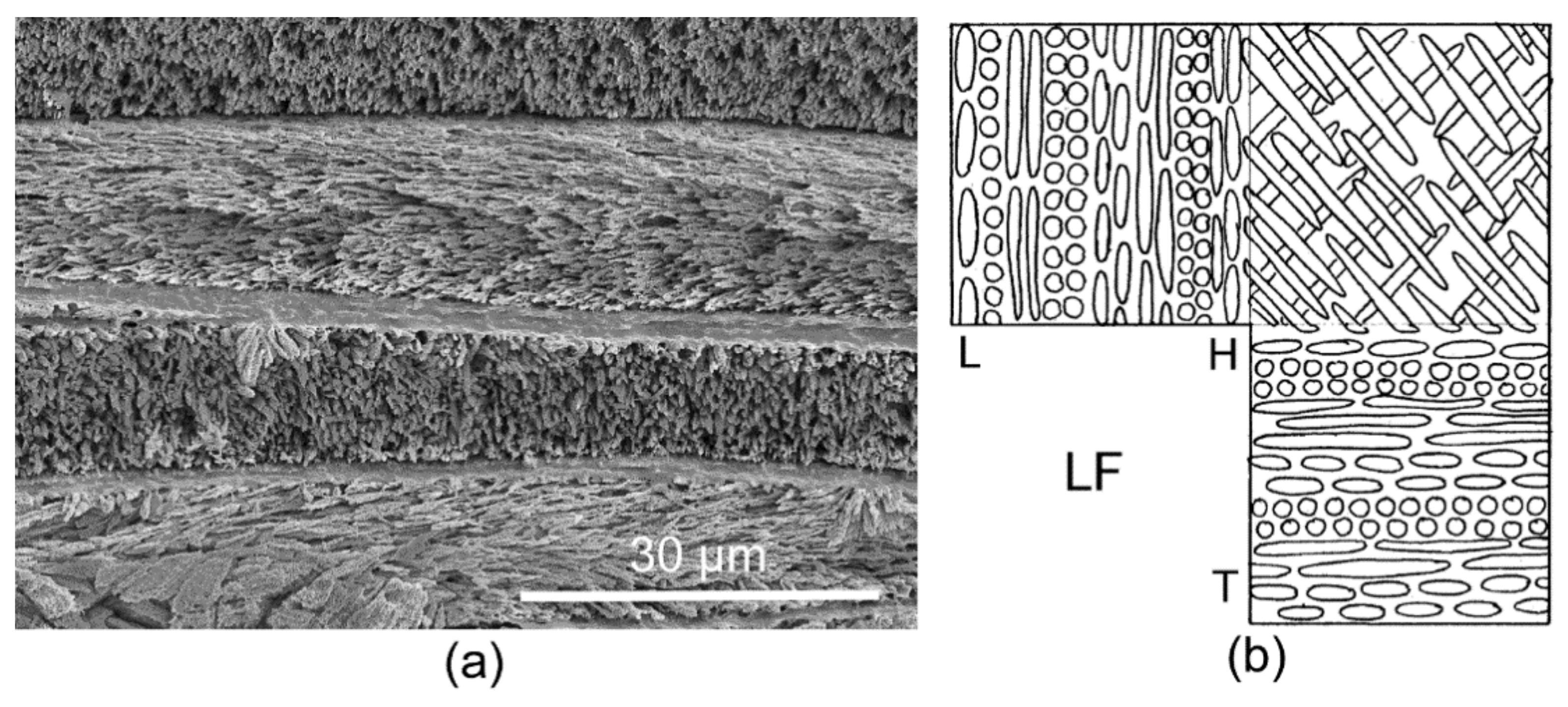
Figure 5.
LF structure in the tube of Cruzigera websteri. (a) Transverse section, inner part of the tube, polished and etched with acetic acid. (b) Schematic line drawing of the LF structure.
Figure 5.
LF structure in the tube of Cruzigera websteri. (a) Transverse section, inner part of the tube, polished and etched with acetic acid. (b) Schematic line drawing of the LF structure.

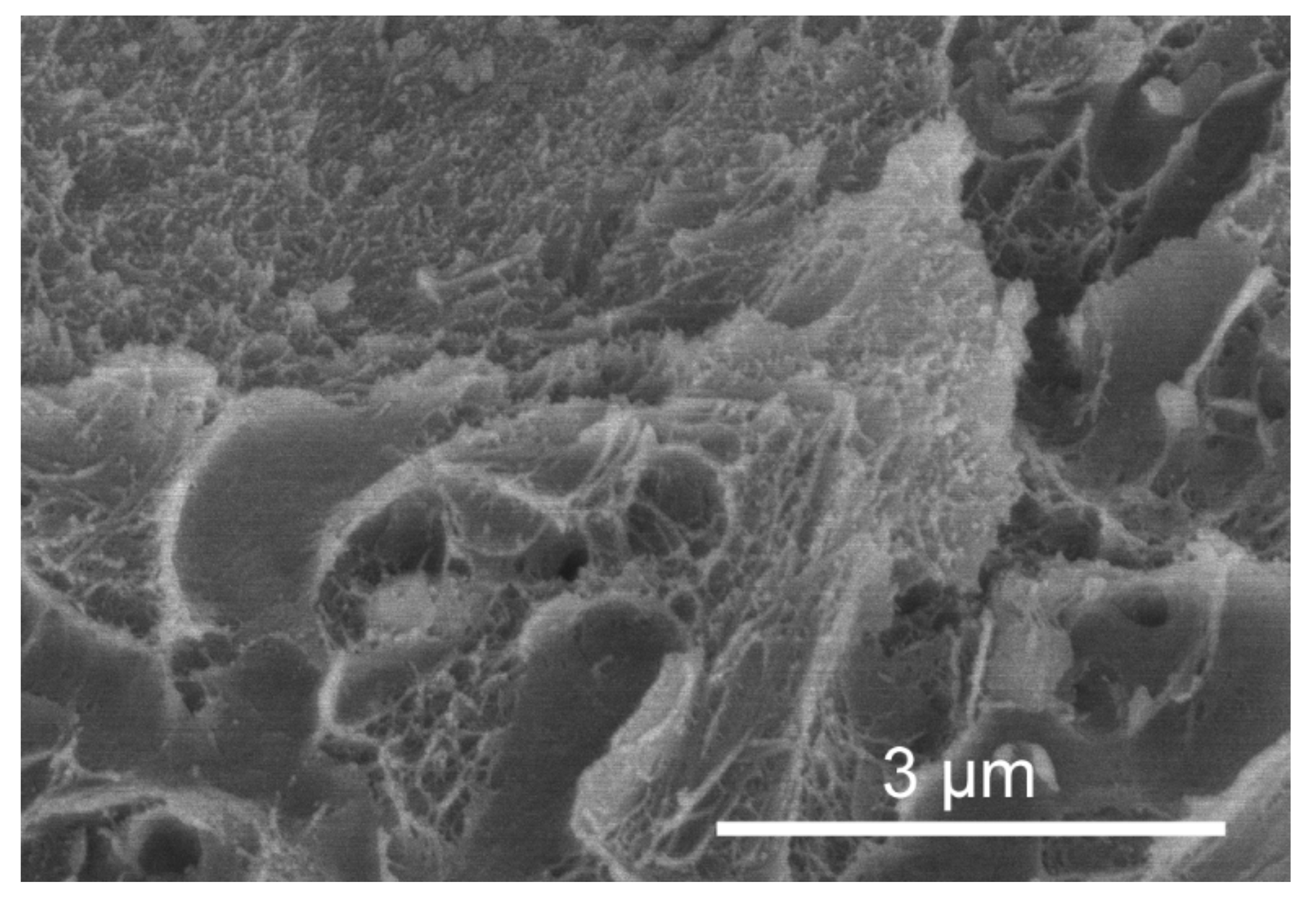
Figure 6.
Inner organic tube lining in Spirobranchus giganteus. Horizontal view.

Figure 7.
Longitudinal section through the tube of Glomerula piloseta. SPHP—spherulitic prismatic structure (inner layer). Orgsh—organic sheets. Sph—spherulites (external layer).
Figure 7.
Longitudinal section through the tube of Glomerula piloseta. SPHP—spherulitic prismatic structure (inner layer). Orgsh—organic sheets. Sph—spherulites (external layer).

Figure 8.
Longitudinal section through the tube of Dodecaceria caulleryi. Spherulitic prismatic structure.
Figure 8.
Longitudinal section through the tube of Dodecaceria caulleryi. Spherulitic prismatic structure.

Figure 9.
Tube structure of Hydroides dianthus. The same growth increment goes across three different microstructures.
Figure 9.
Tube structure of Hydroides dianthus. The same growth increment goes across three different microstructures.

Figure 10.
Evolution of serpulid tube microstructures. Phylogenetic relationships of serpulid genera derived from Kupriyanova et al. [67]. Numbers indicate: 1, unoriented structures; 2, semi-oriented structures; 3, oriented prismatic structures. Serpulids with dominantly calcitic mineralogy or with mixed composition are marked with yellow, and genera with dominantly aragonitic mineralogy are marked with blue.

Publisher’s Note: MDPI stays neutral with regard to jurisdictional claims in published maps and institutional affiliations. |
© 2021 by the author. Licensee MDPI, Basel, Switzerland. This article is an open access article distributed under the terms and conditions of the Creative Commons Attribution (CC BY) license (https://creativecommons.org/licenses/by/4.0/).
Share and Cite
MDPI and ACS Style
Vinn, O. Biomineralization in Polychaete Annelids: A Review. Minerals 2021, 11, 1151. https://doi.org/10.3390/min11101151
AMA Style
Vinn O. Biomineralization in Polychaete Annelids: A Review. Minerals. 2021; 11(10):1151. https://doi.org/10.3390/min11101151
Chicago/Turabian StyleVinn, Olev. 2021. "Biomineralization in Polychaete Annelids: A Review" Minerals 11, no. 10: 1151. https://doi.org/10.3390/min11101151
Note that from the first issue of 2016, this journal uses article numbers instead of page numbers. See further details here.