
Parasites of Free-Ranging and Captive American Primates: A Systematic Review
by
 , , ,
, , ,
Silvia Rondón
1,* ,
,
Serena Cavallero
1,
Erika Renzi
1,
Andrés Link
2,
Camila González
3 and
Stefano D’Amelio
1 1
Department of Public Health and Infectious Diseases, Sapienza University of Rome, Piazzale Aldo Moro 5, 00185 Rome, Italy
2
Laboratorio de Ecología de Bosques Tropicales y Primatología, Departamento de Ciencias Biológicas, Universidad de Los Andes, Cra. 1 N° 18a-12, Bogotá 111711, Colombia
3
Centro de Investigaciones en Microbiología y Parasitología Tropical, CIMPAT, Departamento de Ciencias Biológicas, Universidad de los Andes, Cra. 1 N° 18a-12, Bogotá 111711, Colombia
*
Author to whom correspondence should be addressed.
Microorganisms 2021, 9(12), 2546; https://doi.org/10.3390/microorganisms9122546
Submission received: 14 October 2021
/
Revised: 17 November 2021
/
Accepted: 6 December 2021
/
Published: 9 December 2021
(This article belongs to the Special Issue Molecular Epidemiology and Diagnosis of Parasitic Zoonosis)
Abstract
:The diversity, spread, and evolution of parasites in non-human primates (NHPs) is a relevant issue for human public health as well as for NHPs conservation. Although previous reviews have recorded information on parasites in NHPs (Platyrrhines) in the Americas, the increasing number of recent studies has made these inventories far from complete. Here, we summarize information about parasites recently reported in Platyrrhines, attempting to build on earlier reviews and identify information gaps. A systematic literature search was conducted in PubMed, ISI Web of Science, and Latin American and Caribbean Health Sciences Literature (LILACS), and following the Preferred Reporting Items for Systematic Reviews and Meta-analyses (PRISMA) guidelines. Ninety-three studies were included after the screening process. Records for 20 genera of NHPs, including 90 species were found. Most of the studies were conducted on captive individuals (54.1%), and morphological approaches were the most used for parasite identification. The most commonly collected biological samples were blood and stool, and Protozoa was the most frequent parasite group found. There is still scarce (if any) information on the parasites associated to several Platyrrhine species, especially for free-ranging populations. The use of molecular identification methods can provide important contributions to the field of NHPs parasitology in the near future. Finally, the identification of parasites in NHPs populations will continue to provide relevant information in the context of pervasive habitat loss and fragmentation that should influence both human public health and wildlife conservation strategies.
1. Introduction
Public health, animal welfare, and pathogen transfer to and from wild populations are among the current primary issues of concern in the framework of the One-Health concept. Such aspects are even more relevant in areas of the world such as South America, where biodiversity is declining at high rates and the rate of deforestation is growing. There is compelling evidence on how habitat loss and fragmentation may favor contact between humans and other animals, representing a potential threat for both [1]. In this scenario, non-human primates (NHPs) are of particular interest because of their close phylogenetic relationship with humans and their known role as reservoirs of zoonotic agents [2].
So far, six major groups of organisms have been found infecting NHPs: viruses, bacteria, fungi, protozoa, helminths, and arthropods [3]. For a series of multiple issues including behavioral ecology, public health, and NHPs conservation, it is important to understand the diversity, spread, and evolution of parasites in wild NHPs [4]. Despite this, the inventory of parasites infecting American NHPs is far from complete, highlighting the need for interdisciplinary studies aiming to determine and treat NHPs parasites [5].
Among mammals, the Order Primates includes a high number of species classified as threatened according to the International Union for Conservation of Nature (IUCN), and specifically in Latin America 9% of NHPs species are considered as critically endangered, 12.4% as endangered, and 20.3% as vulnerable according to the Red List [6]. Habitat loss and forest fragmentation are some of the main threats to NHPs species [7], while livestock and ranching are secondary threats affecting 59% of NHPs species in the Neotropics [8].
The implementation of effective measures to reverse anthropogenic pressures against NHPs populations has been strongly encouraged to avoid the imminent loss of NHPs taxa, due to factors such as fragmented landscapes, habitat loss, and degradation, as well as human and domestic animal-borne diseases [8]. As infectious diseases negatively impact NHPs populations, it becomes necessary to watch over all possible introductions of disease, and to better investigate the correlation between diversity and disease exposure risk in humans and wildlife [9]. According to the World Organisation for Animal Health (OIE), the probability of carrying zoonotic pathogens is related to the taxonomic position (increasing from lemurs and tarsiers to marmosets, tamarins and other American monkeys, and finally African and Asian monkeys and apes) and to the region of origin of the species of concern [10]. Likewise, the risk of zoonotic infections involving NHPs is of public health concern, as the expanding human–domestic animal–wildlife interface provides multiple opportunities for the agents of disease to shift hosts. Additionally, fragmentation and habitat loss interrupt natural processes involving parasites and hosts [11].
In this context, molecular epidemiology and diagnosis of parasitic zoonoses in NHPs play a very important role in the understanding of parasite ecology and the assessment of their zoonotic potential. Previous reviews have gathered information of parasites in Platyrrhines [5,12] or have been focused on the fragmentation of the fauna living in the American tropics [13,14]. Given the large number of recent studies, it is worth updating and compiling NHPs parasitological information along with information related to threatening factors, creating a useful tool that may serve as the basis to better direct future research projects, support decision making and fill information gaps.
This systematic review summarizes information about parasites (protozoans, helminths and ectoparasites) recently reported in American NHPs, building on previous reviews and attempting to identify information gaps regarding the parasitic pathogens circulating in a major concern group of hosts and in a critical biodiverse region.
2. Materials and Methods
We carried out a systematic review following the Preferred Reporting Items for Systematic Reviews and Meta-Analyses (PRISMA) to summarize information about parasites infecting American NHPs. The review protocol of this systematic review was not recorded into the International prospective register of systematics reviews (PROSPERO) (Supplementary File S2: Authors declaration and PRISMA checklist). We performed an independent search for each Platyrrhine genus, using the terms “parasite” and NHPs genus (e.g., parasite AND Cebus). The search was conducted in ISI Web of Knowledge and PubMed, including studies from June 2017 to 11 February 2021, thus collecting all the information published after the time frame used in the last available review regarding the subject [5]. Additionally, information from Latin American and Caribbean Health Sciences Literature (LILACS) was incorporated into the database, using the same search terms, until February 11th, 2021. In this way we collected information from a Latin American specific search engine, building on the review made by Solórzano-García and Pérez-Ponce de León [5].
We included studies performed in wild and captive Platyrrhines which reported parasite occurrence, while studies under laboratory conditions or the ones focused on fungi, bacteria, and viruses were not included. We used articles in English, Portuguese, or Spanish.
Two reviewers screened the records independently. In case of a disagreement that was not consensually solved, a third reviewer arbitrated the decision process. For data extraction, we used a standardized form that included the following features: host family, host genera, host species, collected sample (stool, blood, tissue, ectoparasite), parasite detection method (PCR, microscopy, etc.), parasite family, parasite genus, parasite species, parasite prevalence (%), parasite group (protozoa, cestoda, nematoda, trematoda, phthiraptera, acariformes, ixodida, diptera, pentastomida, siphonaptera), endoparasite/ectoparasite, forest fragmentation evaluated (yes/no), country, state and habitat (sylvatic/captivity).
To organize the collected data, we considered specific taxonomy classifications: for Lagothrix, Saguinus, and Callicebus, we followed the classification proposed by Di Fiore et al. [15], Buckner et al. [16], and Byrne et al. [17], respectively. For all other NHPs genera, we followed the taxonomy of the “Handbook of the mammals of the world” [18]. Parasite taxonomy was included following the classification stated by the National Center for Biotechnology Information (NCBI).
3. Results
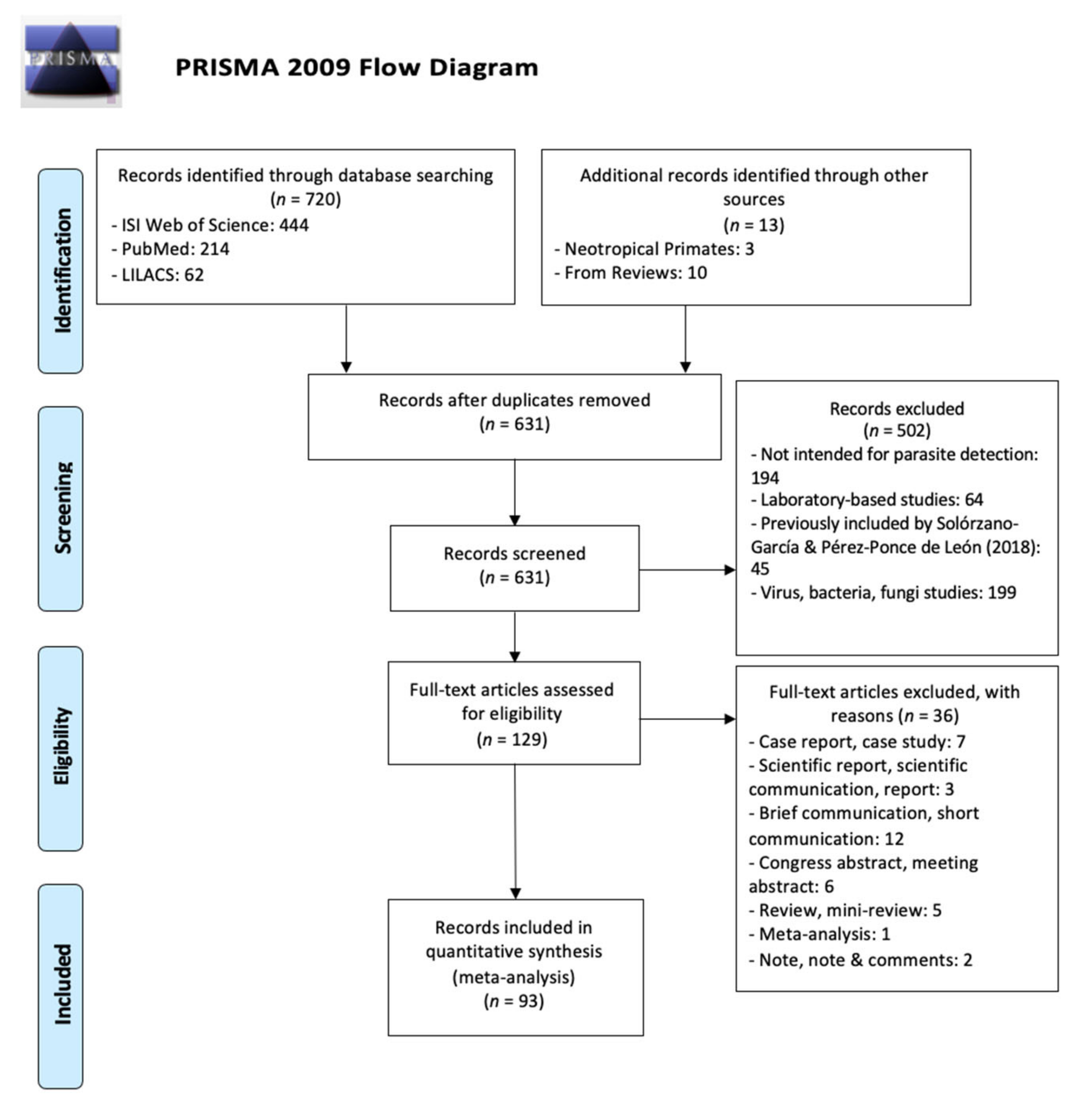
The literature review retrieved 720 searches: 444 from ISI Web of Knowledge, 214 from PubMed, and 62 from LILACS. Overall, we obtained 93 novel publications (Figure 1) after eliminating duplicates, studies under laboratory conditions, studies already included in the review made by Solórzano-García & Pérez-Ponce de León [5].
Overall, the studies included in this review account for 20 Platyrrhine genera, including 90 species. The genus with most records was Alouatta (n = 51), while genera with the least records were Callimico (n = 1) and Cebuella (n = 1) (Table 1). According to the parasite group, protozoa were overall the most reported along NHPs genera (Table 1). It was found that 54.1% studies were conducted on captive NHPs and 45.9% on free-ranging animals, while the source of biological sample and diagnostic method mostly used were blood and morphology, respectively (Table 2).
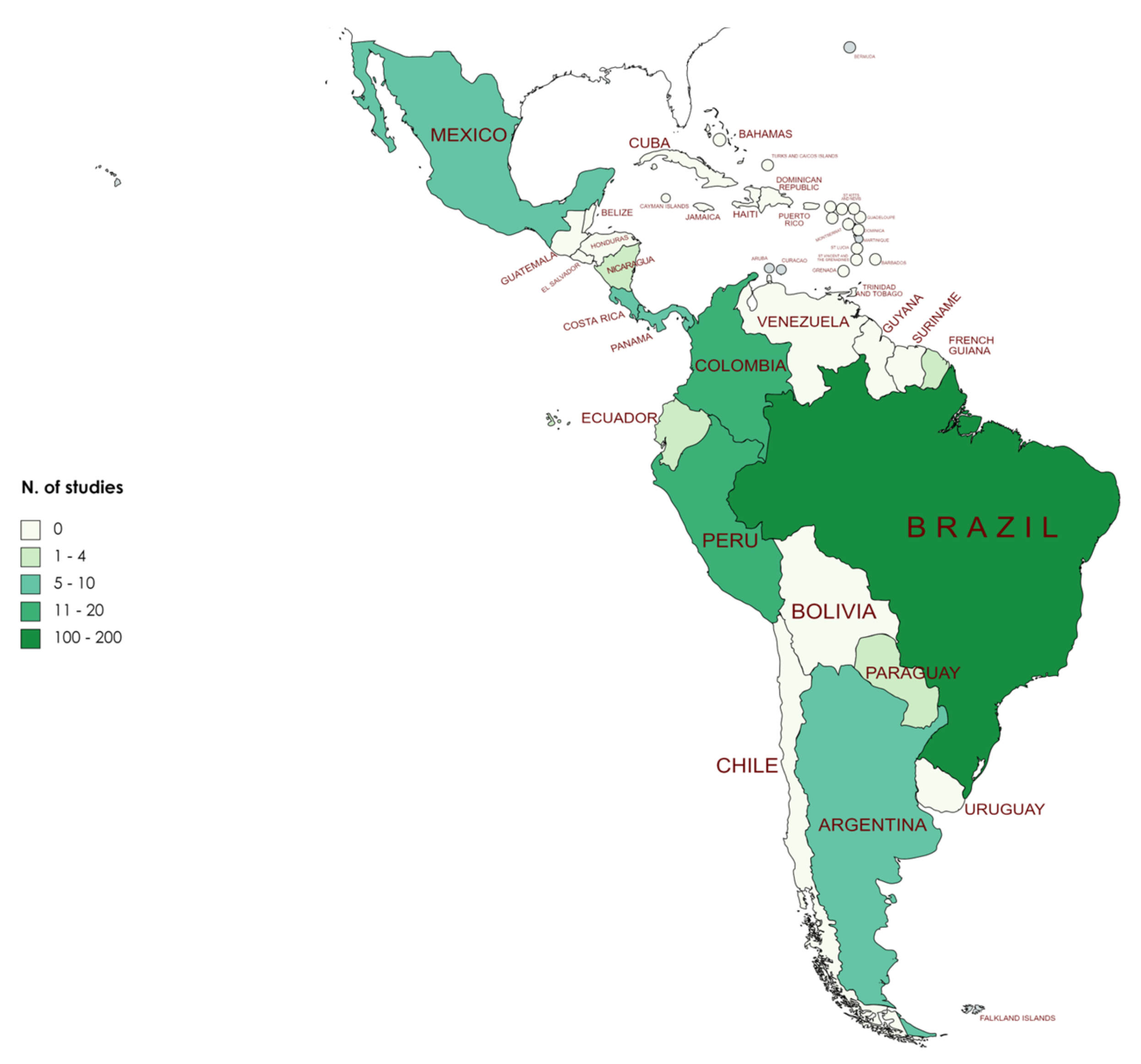
A list of parasites per NHPs species is shown (Table 3), as well as a list of parasite-host (Supplementary File S1). When considering the geographical distribution of the records, Brazil was the country with most of them, including information for 19 NHPs genera. There were no publications for Belize, Bolivia, Guatemala, Guyana, Honduras, El Salvador, Suriname, and Venezuela (Figure 2). Additionally, there were publications regarding captive Neotropical NHPs in Europe and Asia: Aotus, Callimico, and Cebuella in Switzerland (1 record each), Cebus in France (1 record), Saguinus in Italy (1 record), Ateles, Saimiri, and Sapajus in China (1, 2, and 1 records, respectively), Callithrix in Korea (1 record), and Saimiri in Japan and South Korea (1 record for each country).
Alouatta was the NHPs genus with most records (n = 51) followed by Callithrix (n = 22), while for all other genera less than 20 records were retrieved (Table 1). Considering the number of species, the genera with more species are Plecturocebus (n = 23), Saguinus (n = 15), Cebus (n = 14), Alouatta (n = 12), and Aotus (n = 11). Likewise, Ateles, Brachyteles, Callimico, Callithrix, Cebuella, and Sapajus had records for 100% of the species, and 80% of the species for Lagothrix, Pithecia, and Saguinus. Brazil was the country with most parasitological records (n = 163), being also the country with highest recorded occurrence of NHPs. Other NHPs rich-countries, such as Peru and Colombia, were the second and third countries with more records (20 and 13, respectively).
4. Discussion
The most recent list (2018–2020) of the World’s 25 Most Endangered NHPs Species includes six Platyrrhines: Ateles geoffroyi, Cebus aequatorialis, Saguinus bicolor, Plecturocebus olallae, Alouatta guariba, and Callithrix aurita [112]. After the systematic review process, there were retrieved publications with parasitological data for A. geoffroyi (n = 4), S. bicolor (n = 4), A. guariba (n = 11), and C. aurita (n = 1), while there were no articles mentioning C. aequatorialis and P. olallae. Additionally, there were no records for Plecturocebus caquetensis, P. olallae, Leontopithecus caissara, and Callicebus barbarabrownae, listed in the IUCN Red List as Critically Endangered [6], neither for Cebus malitiosus, Saimiri vanzolinii, Callicebus coimbrai, Alouatta ululata, or Cebus cesarae, listed as Endangered [6]. The amount of information is probably biased by the availability of different species in captivity, a condition that strongly facilitate parasitological investigations. It can be speculated that the lack of information for endangered species could be related to their scarcity in captive conditions. Although other kinds of studies (e.g., behavioral, genetic) may have been carried out for those species during the time range considered in this study, it must be highlighted that parasitological studies are also very important, representing a useful insight for monitoring the health status of NHPs in contexts of human–NHPs interfaces, as human-induced forest loss increase the exposition of NHPs to human and domesticated animal pathogens [8]. Additionally, even if non-lethal parasite infections are common in wild NHPs, parasite infections could cause sickness behaviors that may be adaptative in the short-term but have longer-term fitness consequences [113]. Note that for some Critically Endangered and Endangered NHPs species which had no parasitological studies until 2017, data have been recorded between 2017 and 2021, as is the case of Cebus kaapori, Sapajus flavius, and Ateles marginatus. Moreover, even if there are reports for specific NHPs species, the observation is limited to a specific area implying that not all the geographic range of the species has been covered.
Overall, just over half of the studies were conducted on captive NHPs (54.1%), however, for the genera Alouatta, Cacajao, Callithrix, and Leontocebus there were more records on free-ranging NHPs. Studies in both free-ranging and captive NHPs are important, for instance, in the design of conservation strategies, reintroduction programs, and NHPs acquisition for research laboratories or zoos. Determining the composition of parasite communities in captive NHPs allows the identification of parasites of concern regarding the introduction of novel parasites to potentially susceptible wildlife populations during reintroduction programs, and also lead to a better understand parasite ecology, for instance, it has been observed that vector-borne parasites are more likely found in free-ranging NHPs, while parasites transmitted through either close and non-close contact, including the fecal–oral transmission, are more likely detected in captive NHPs [114].
Regarding the diagnostic method, morphological approaches were found to be the most used, followed by molecular procedures.
The most common biological samples were blood and stool, and ectoparasites corresponded to the least reported. Sampling NHPs, specially free-ranging, is logistically challenging as invasive sampling techniques such as the collection of blood, requires field anesthesia; therefore, optimization of non-invasive surveillance on NHPs is critical for understanding disease ecology of pathogens and identifying zoonotic diseases likely to emerge [115]. In this context, non-invasive methods such as stool collection are among the safest alternatives to study multiple aspects of the biology of NHPs [2]. However, even the collection of stool samples requires considerable efforts for their assignment to a specific individual, as well as to avoid multiple sampling for the same individual and later calculate the prevalence of parasites.
Parasitological surveys of NHPs contribute to the understanding of the epidemiology, zoonotic emergence risk and transmission dynamics [41]. In this context, parasitological studies using adequate tools to evaluate the zoonotic potential are necessary. In the present review, as most studies are based on parasite morphology, some parasite species and/or genetic variants could not be determined, thus not allowing to assess their zoonotic potential. In future studies, the use of molecular tools will become essential, not only to identify and determine the presence/absence of parasites, but also to identify species/variants of the parasites circulating in each NHPs species and in each sampling site in order to better understand their distribution in NHPs and to evaluate transmission dynamics. Although there are challenges related to the molecular processing of the samples (e.g., disruption of the Ascaris and Trichuris eggshells prior to DNA extraction), efforts should be made to develop efficient protocols especially in stool samples. These are considered reliable for the non-invasive detection of pathogens, opening up new possibilities in the molecular epidemiology and evolutionary analysis of infectious diseases [2].
Molecular tools have been mainly used in studies aimed to detect protozoans: Plasmodium sp., Toxoplasma sp., Entamoeba sp., Giardia sp., Blastocystis sp., Leishmania sp., Trypanosoma sp., and Pentatrichomonas sp. [25,42,45,64,65,76,85]. However, the determination of parasite subtypes/genetic lineages was performed in few studies. Some studies on Blastocystis assessed the genetic variability and host specificity, reporting different subtypes (ST1-ST5, ST8) [42,61,116]. Studies on Trypanosoma cruzi identified the genetic lineages of the parasite (TcI-TcIII, TcV, TcVI) [64,90] as well as types of Toxoplasma gondii (Type I, Type II, non-archetypal) [39,100]. Molecular approaches were less frequent in studies of nematodes: Trypanoxyuris sp., Dipetalonema sp., Mansonella sp., Brugia sp., Pterygodermatites sp. [26,59,102,117], cestodes: Mesocestoides sp. [96], and ectoparasites: Amblyomma sp. [109]. No molecular records on zoonotic parasites as Trichuris sp., Ascaris sp., Cryptosporidium sp., Hymenolepis sp., Taenia sp., Strongyloides sp., Capillaria sp., nor Balantidium sp. were found.
As habitat loss and forest fragmentation are currently a concerning global trend, and NHPs are in closer contact with humans, consequent ecological changes need to be monitored. Forest fragmentation is one of the main factors threatening NHPs [8], affecting all but not only the six Neotropical species included into the World’s 25 Most Endangered NHPs List [112]. However, only two studies included in the present review accounted for forest fragmentation as a variable during the analyses [25,45], even if some were performed in fragmented areas [67,81,85,91,105]. We strongly encourage the inclusion of this crucial factor as a variable for future studies as a way of better understanding parasite ecology, taking into account that some studies had reported a higher parasite prevalence in NHPs living in fragmented habitats [62,118], while other authors had found a lower presence of parasites [69], in comparison to prevalence found in continuous forests. Additionally, parasite taxa composition may vary according to NHPs living condition [118,119].
Not only more efforts aimed to broaden the knowledge of parasites infecting NHPs are required but we also suggest the standardization of the result presentation/display. For instance, it is necessary to include the coordinates of the sampling sites and show information separately for each NHPs species and study site when sampling is simultaneously carried out in different sites, involving more than one NHPs species. Therefore, the availability of the necessary information to perform meta-analysis, spatial analyses, and calculate parasite prevalence is facilitated, allowing to draw conclusions usable to a better understanding of infection patterns.
5. Conclusions
In the present review, parasitological records for 20 genera of NHPs mainly conducted on captive animals were retrieved. Morphological approaches were found to be the most used, and Protozoa was the most frequent parasite group reported. Parasitological studies on American NHPs still need to be performed, especially for some genera and species with several information gaps, as well as Critically Endangered and Endangered primates, in both free-ranging and captive conditions. Parasitological studies using adequate tools to evaluate potential zoonoses are necessary in order to better understand the distribution of parasites in NHPs and to evaluate transmission dynamics, also taking considering factors as habitat loss and forest fragmentation.
Supplementary Materials
The following are available online at https://www.mdpi.com/article/10.3390/microorganisms9122546/s1: Supplementary File S1: Parasite-host list, Supplementary File S2: Authors declaration regarding the pre-recorded protocol.
Author Contributions
Conceptualization: S.R., C.G., A.L. and S.C.; literature review: S.R. and E.R.; PRISMA guidelines: E.R.; primate taxonomy corrections: A.L.; supervision of the project: S.D. and S.C.; writing—review and editing: all authors. All authors have read and agreed to the published version of the manuscript.
Funding
This research received no external funding.
Institutional Review Board Statement
Not applicable.
Informed Consent Statement
Not applicable.
Data Availability Statement
Not applicable.
Acknowledgments
We thank the authors who have shared useful information or details for the development of the database generated in this review. We also thank the anonymous reviewers of the manuscript.
Conflicts of Interest
The authors declare no conflict of interest.
References
- Goldberg, T.L.; Gillespie, T.R.; Rwego, I.B.; Estoff, E.L.; Chapman, C.A. Forest fragmentation as cause of bacterial transmission among nonhuman primates, humans, and livestock, Uganda. Emerg. Infect. Dis. 2008, 14, 1375–1382. [Google Scholar] [CrossRef]
- Davoust, B.; Levasseur, A.; Mediannikov, O. Studies of nonhuman primates: Key sources of data on zoonoses and microbiota. New Microbes New Infect. 2018, 26, S104–S108. [Google Scholar] [CrossRef] [PubMed]
- Nunn, C.; Altizer, S. Diversity and characteristics of primate parasites. In Infectious Diseases in Primates-Behaviour, Ecology and Evolution; Oxford University Press: New York, NY, USA, 2006; p. 26. [Google Scholar]
- Nunn, C.; Altizer, S. The global mammal parasite database: An online resource for infectious disease records in wild primates. Evol. Anthropol. Issues News Rev. 2005, 14, 1–2. [Google Scholar] [CrossRef]
- Solórzano-García, B.; Pérez-Ponce de León, G. Parasites of Neotropical Primates: A Review. Int. J. Primatol. 2018, 39, 155–182. [Google Scholar] [CrossRef]
- IUCN Primates Order- Mesoamerica & South America-Species. Available online: https://www.iucnredlist.org/search/stats?query=primates&searchType=species (accessed on 9 June 2021).
- Bicca-Marques, J.; Chaves, Ó.; Hass, G. Howler monkey tolerance to habitat shrinking: Lifetime warranty or death sentence? Am. J. Primatol. 2020, 4, e23089. [Google Scholar] [CrossRef]
- Estrada, A.; Garber, P.; Rylands, A.; Roos, C.; Fernandez-Duque, E.; Di Fiore, A.; Nekaris, K.; Nijman, V.; Heymann, E.; Lambert, J.; et al. Impending extinction crisis of the world’s primates: Why primates matter. Sci. Adv. 2017, 3, e1600946. [Google Scholar] [CrossRef] [PubMed] [Green Version]
- Nunn, C.; Gillespie, T. Infectious disease and primate conservation. In An introduction to Primate Conservation; Wich, S., Marshall, A., Eds.; Oxford University Press: New York, NY, USA, 2016; p. 168. [Google Scholar]
- OIE World Organisation for Animal Health Zoonoses Transmissible from Non-Human Primates. OIE Terrestrial Animal Health Code; OIE: Paris, France, 2019; Volume 1. [Google Scholar]
- Frias, L.; MacIntosh, J. Threatened Hosts, Threatened Parasites?-Parasite Diversity and Distribution in Red-Listed Primates. In Primate Research and Conservation in the Anthropocene; Behie, A., Teichroeb, J., Malone, N., Eds.; Cambridge University Press: Cambridge, UK, 2019; p. 142. [Google Scholar]
- Corrêa, P.; Bueno, C.; Soares, R.; Vieira, F.; Muniz-Pereira, L. Checklist of helminth parasites of wild primates from Brazil. Rev. Mex. Biodivers. 2016, 87, 908–918. [Google Scholar] [CrossRef] [Green Version]
- Arroyo-Rodríguez, V.; Dias, P. Effects of habitat fragmentation and disturbance on howler monkeys: A review. Am. J. Primatol. 2010, 72, 1–16. [Google Scholar] [CrossRef]
- Offerman, H.; Dale, V.; Pearson, S.; Bierregaard, R.; O’Neill, R. Effects of forest fragmentation on neotropical fauna: Current research and data availability. Environ. Rev. 1995, 3, 191–211. [Google Scholar] [CrossRef]
- Di Fiore, A.; Chaves, P.B.; Cornejo, F.M.; Schmitt, C.A.; Shanee, S.; Cortés-Ortiz, L.; Fagundes, V.; Roos, C.; Pacheco, V. The rise and fall of a genus: Complete mtDNA genomes shed light on the phylogenetic position of yellow-tailed woolly monkeys, Lagothrix flavicauda, and on the evolutionary history of the family Atelidae (Primates: Platyrrhini). Mol. Phylogenet. Evol. 2015, 82, 495–510. [Google Scholar] [CrossRef] [PubMed]
- Buckner, J.C.; Lynch Alfaro, J.W.; Rylands, A.B.; Alfaro, M.E. Biogeography of the marmosets and tamarins (Callitrichidae). Mol. Phylogenet. Evol. 2015, 82, 413–425. [Google Scholar] [CrossRef] [PubMed]
- Byrne, H.; Rylands, A.B.; Carneiro, J.C.; Alfaro, J.W.L.; Bertuol, F.; da Silva, M.N.F.; Messias, M.; Groves, C.P.; Mittermeier, R.A.; Farias, I.; et al. Phylogenetic relationships of the New World titi monkeys (Callicebus): First appraisal of taxonomy based on molecular evidence. Front. Zool. 2016, 13. [Google Scholar] [CrossRef] [PubMed] [Green Version]
- Wilson, D.E.; Mittermeier, R.A. (Eds.) Handbook of the Mammals of the World, Volume 3—Primates; Lynx Edicions: Barcelona, Spain, 2014; ISBN 978-84-96553-89-7. [Google Scholar]
- De la Hoz, D.M.E.; Cañate González, A.S.; Vergel, E.F.; Payares Ramírez, K.J.; Morales López, S.E. Parasitic and fungal agents in Aotus sp., Alouatta seniculus and Cebus albifrons in the Colombian Caribbean. Rev. Investig. Vet. Peru 2020, 31. [Google Scholar] [CrossRef]
- Bouer, A.; Werther, K.; Zacarias, R.; Higa, A.; Epiphanio, S.; Catão-Dias, J. Detection of anti-Toxoplasma gondii antibodies in experimentally and naturally infected non-human primates by Indirect Fluorescence Assay (IFA) and indirect ELISA. Revista Brasileira de Parasitologia Veterináriá 2010, 19, 26–31. [Google Scholar] [CrossRef] [PubMed] [Green Version]
- Grumann, M.R.; Da Silva, Z.; Filho, J.R.S.; Costa, M.M.; Vieira, M.I.B.; Da Motta, A.C. Immunohistochemical and serological aspects of Toxoplasma gondii infection in Neotropical primates. Semin. Agrar. 2017, 38, 1375–1382. [Google Scholar] [CrossRef]
- Aysanoa, E.; Mayor, P.; Mendoza, A.P.; Zariquiey, C.M.; Morales, E.A.; Pérez, J.G.; Bowler, M.; Ventocilla, J.A.; González, C.; Baldeviano, G.C.; et al. Molecular Epidemiology of Trypanosomatids and Trypanosoma cruzi in Primates from Peru. Ecohealth 2017, 14, 732–742. [Google Scholar] [CrossRef] [PubMed]
- Deane, L.M. Simian malaria in Brazil. Memórias do Instituto Oswaldo Cruz 1992, 87, 1–20. [Google Scholar] [CrossRef]
- Bahia, M.; de Nazaré Leite Barros, F.; Magalhães-Matos, P.C.; de Souza Gonçalves, T.; Chiesorin Neto, L.; Oliveira Faria, D.C.L.; Aparecida Romeiro, S.; Barros Monteiro, F.O.; Góes-Cavalcante, G.; Scofield, A. Trypanosoma cruzi infection in captive Neotropical primates in the Brazilian Amazon. Am. J. Primatol. 2017, 79, e22590. [Google Scholar] [CrossRef] [PubMed]
- Rondón, S.; León, C.; Link, A.; González, C. Prevalence of Plasmodium parasites in non-human primates and mosquitoes in areas with different degrees of fragmentation in Colombia. Malar. J. 2019, 18, 1–10. [Google Scholar] [CrossRef]
- Balsiger, A.; Federer, K.; Grimm, F.; Deplazes, P. Transmission of Pterygodermatites nycticebi in a colony of goeldi’s monkeys (Callimico goeldii) and evaluation of treatment and control. J. Zoo Wildl. Med. 2018, 49, 893–901. [Google Scholar] [CrossRef]
- Figueiredo, M.; Di Santi, S.; Gómez, W.; André, M.; Zacarias, R. Identification of Plasmodium spp. in Neotropical primates of Maranhense Amazon in Northeast Brazil. PLoS ONE 2017, 12, e0182905. [Google Scholar] [CrossRef] [Green Version]
- Figueiredo, M.A.P.; Di Santi, S.M.; Manrique, W.G.; André, M.R.; Machado, R.Z. Serological and molecular techniques applied for identification of Plasmodium spp. in blood samples from nonhuman primates. Rev. Bras. Parasitol. Vet. 2018, 27, 363–376. [Google Scholar] [CrossRef] [PubMed] [Green Version]
- Lombardi, M.C.; Turchetti, A.P.; Tinoco, H.P.; Pessanha, A.T.; Soave, S.A.; Malta, M.C.C.; Paixão, T.A.; Santos, R.L. Diagnosis of Leishmania infantum infection by polymerase chain reaction in wild mammals. Pesqui. Vet. Bras. 2014, 34, 1243–1246. [Google Scholar] [CrossRef] [Green Version]
- Santos, R.; Oliveira, A. Leishmaniasis in non-human primates: Clinical and pathological manifestations and potential as reservoirs. J. Med. Primatol. 2020, 49, 34–39. [Google Scholar] [CrossRef] [PubMed]
- Roncancio, N.; Santa, M.A.; Calderón, L.M.; Gómez, E.N.; Acosta, A.; García, L.M.; Henao, B.E.; Peñuela, S.M.; Pinilla, E.A.; Poches, R.A.; et al. Differences in the prevalence of cutaneous myiasis between Aotus vociferans and Aotus nancymaae in the Colombian Amazon. Neotrop. Primates 2018, 24, 86–90. [Google Scholar]
- Paula, N.F.D.; Dutra, K.S.; Oliveira, A.R.D.; Santos, D.O.D.; Rocha, C.E.V.; Vitor, R.W.D.A.; Tinoco, H.P.; Costa, M.E.L.T.D.; Paixão, T.A.D.; Santos, R.L. Host range and susceptibility to Toxoplasma gondii infection in captive neotropical and Old-world primates. J. Med. Primatol. 2020, 49, 202–210. [Google Scholar] [CrossRef] [PubMed]
- Rodrigues de Oliveira, A.; Pinheiro, G.; Tinoco, H.; Loyola, M.; Coelho, C.; Dias, E.; Monteiro, E.; de Oliveira Lara e Silva, F.; Tinoco Pessanha, A.; Souza, A.; et al. Competence of non-human primates to transmit Leishmania infantum to the invertebrate vector Lutzomyia longipalpis. PLoS Negl. Trop. Dis. 2019, 13, e0007313. [Google Scholar] [CrossRef] [PubMed]
- Helenbrook, W.D.; Nelson, A.; Paras, K.L.; Solorzano-Garcia, B. Intestinal Parasitism in Free-Ranging Black-Headed Night Monkeys, Aotus nigriceps, of Southeastern Peru. Int. J. Primatol. 2020, 41, 458–470. [Google Scholar] [CrossRef]
- Chagas, C.R.F.; Gonzalez, I.H.L.; Salgado, P.A.B.; Rodrigues, B.; Ramos, P.L. Giardia spp., ten years of parasitological data in the biggest zoo of Latin America. Ann. Parasitol. 2019, 65, 35–51. [Google Scholar] [CrossRef]
- Herrer, A.; Christensen, H. Epidemiological patterns of cutaneous leishmaniasis in Panama III. Endemic persistence of the disease. Am. J. Trop. Med. Hyg. 1976, 25, 54–58. [Google Scholar] [CrossRef]
- Ramírez, J.D.; Sánchez, L.V.; Bautista, D.C.; Corredor, A.F.; Flórez, A.C.; Stensvold, C.R. Blastocystis subtypes detected in humans and animals from Colombia. Infect. Genet. Evol. 2014, 22, 223–228. [Google Scholar] [CrossRef] [PubMed]
- Buery, J.C.; Rodrigues, P.T.; Natal, L.; Salla, L.C.; Loss, A.C.; Vicente, C.R.; Rezende, H.R.; Duarte, A.M.R.D.C.; Fux, B.; Malafronte, R.D.S.; et al. Mitochondrial genome of Plasmodium vivax/simium detected in an endemic region for malaria in the Atlantic Forest of Espírito Santo state, Brazil: Do mosquitoes, simians and humans harbour the same parasite? Malar. J. 2017, 16. [Google Scholar] [CrossRef] [PubMed] [Green Version]
- Santana, C.H.; de Oliveira, A.R.; dos Santos, D.O.; Pimentel, S.P.; de Souza, L.D.R.; Moreira, L.G.A.; Braz, H.M.B.; de Carvalho, T.P.; Lopes, C.E.B.; Oliveira, J.B.S.; et al. Genotyping of Toxoplasma gondii in a lethal toxoplasmosis outbreak affecting captive howler monkeys (Alouatta sp.). J. Med. Primatol. 2020. [Google Scholar] [CrossRef] [PubMed]
- Solórzano-García, B.; Gasca-Pineda, J.; Poulin, R.; Pérez-Ponce de León, G. Lack of genetic structure in pinworm populations from New World primates in forest fragments. Int. J. Parasitol. 2017, 47, 941–950. [Google Scholar] [CrossRef] [PubMed]
- Martínez, M.F.; Kowalewski, M.M.; Giuliani, M.G.; Acardi, S.A.; Salomón, O.D. Molecular identification of Leishmania in free-ranging black and gold howler monkeys (Alouatta caraya) in northeastern Argentina. Acta Trop. 2020, 210. [Google Scholar] [CrossRef]
- Oliveira-Arbex, A.P.; David, É.B.; da Silva Tenório, M.; Cicchi, P.J.P.; Patti, M.; Coradi, S.T.; Lucheis, S.B.; Jim, J.; Guimarães, S. Diversity of Blastocystis subtypes in wild mammals from a zoo and two conservation units in southeastern Brazil. Infect. Genet. Evol. 2020, 78, 104053. [Google Scholar] [CrossRef] [PubMed]
- Milozzi, C.; Bruno, G.; Cundom, E.; Mudry, M.D.; Navone, G.T. Intestinal parasites of Alouatta caraya (Primates, Ceboidea): Preliminary study in semi-captivity and in the wild in Argentina. Mastozoologia Neotrópical 2012, 19, 271–278. [Google Scholar]
- Servián, A.; Zonta, M.L.; Cociancic, P.; Falcone, A.; Ruybal, P.; Capasso, S.; Navone, G.T. Morphological and molecular characterization of Bertiella sp. (Cestoda, Anoplocephalidae) infection in a human and howler monkeys in Argentina. Parasitol. Res. 2020, 119, 1291–1300. [Google Scholar] [CrossRef] [PubMed]
- Kuthyar, S.; Kowalewski, M.M.; Roellig, D.M.; Mallott, E.K.; Zeng, Y.; Gillespie, T.R.; Amato, K.R. Effects of anthropogenic habitat disturbance and Giardia duodenalis infection on a sentinel species’ gut bacteria. Ecol. Evol. 2021, 11, 45–57. [Google Scholar] [CrossRef] [PubMed]
- Duarte, A.M.R.D.C.; Porto, M.A.L.; Curado, I.; Malafronte, R.S.; Hoffmann, E.H.E.; de Oliveira, S.G.S.G.; da Silva, A.M.J.; Kloetzel, J.K.; Gomes, A.D.C. Widespread occurrence of antibodies against circumsporozoite protein and against blood forms of Plasmodium vivax, P. falciparum and P. malariae in Brazilian wild monkeys. J. Med. Primatol. 2006, 35, 87–96. [Google Scholar] [CrossRef]
- Kane, J.; Smith, R.L. Bertiella sp. (Meyner, 1895) infection of Alouatta caraya (Humbolt, 1812) in urban and natural environments in Ñeembucú, southwest Paraguay. Am. J. Primatol. 2020. [Google Scholar] [CrossRef]
- Pereira, F.V.; Lucena, F.P.; Rodrigues, R.L.; Barros, L.A.; Pires, C.A.; Ferreira, A.M.R.; Mello, M.F.V. Prevalence and spatial distribution of the occurrence of helminths in free-living nonhuman primates in the State of Rio de Janeiro, Brazil. Arq. Bras. Med. Vet. Zootec. 2020, 72, 1705–1712. [Google Scholar] [CrossRef]
- Brasil, P.; Zalis, M.G.; de Pina-Costa, A.; Siqueira, A.M.; Júnior, C.B.; Silva, S.; Areas, A.L.L.; Pelajo-Machado, M.; de Alvarenga, D.A.M.; da Silva Santelli, A.C.F.; et al. Outbreak of human malaria caused by Plasmodium simium in the Atlantic Forest in Rio de Janeiro: A molecular epidemiological investigation. Lancet Glob. Health 2017, 5, e1038–e1046. [Google Scholar] [CrossRef] [Green Version]
- De Alvarenga, D.; Culleton, R.; Pina-costa, A.; Fonseca, D.; Junior, C.; Silva, S.; Dutra, A.; de Souza, J.; Braga, Z.; Moreira, S.B.; et al. An assay for the identification of Plasmodium simium infection for diagnosis of zoonotic malaria in the Brazilian Atlantic Forest. Sci. Rep. 2018, 8, 86. [Google Scholar] [CrossRef] [PubMed] [Green Version]
- Da Fonseca, F. Plasmodium of a primate of Brazil. Memórias do Instituto Oswaldo Cruz 1951, 49. [Google Scholar] [CrossRef]
- Abreu, F.V.S.D.; Santos, E.D.; Mello, A.R.L.; Gomes, L.R.; Alvarenga, D.A.M.D.; Gomes, M.Q.; Vargas, W.P.; Bianco-Júnior, C.; Pina-Costa, A.D.; Teixeira, D.S.; et al. Howler monkeys are the reservoir of malarial parasites causing zoonotic infections in the Atlantic forest of Rio de Janeiro. PLoS Negl. Trop. Dis. 2019, 13, e0007906. [Google Scholar] [CrossRef]
- Nunes, A.J.D.; de Alvarenga, D.A.M.; de Souza Junior, J.C.; Peruchi, A.R.; Gonçalves, G.H.P.; Hirano, Z.M.B.; de Brito, C.F.A.; Cremer, M.J. Plasmodium infection and its association with biochemical and haematological parameters in free-living Alouatta guariba clamitans (Cabrera, 1940) (primates: Atelidae) in Southern Brazil. Memórias do Instituto Oswaldo Cruz 2019, 114. [Google Scholar] [CrossRef] [PubMed] [Green Version]
- De Abreu, F.V.S.; Gomes, L.R.; Mello, A.R.L.; Bianco-Júnior, C.; De Pina-Costa, A.; Dos Santos, E.; Teixeira, D.S.; Brasil, P.; Daniel-Ribeiro, C.T.; Lourenço-De-Oliveira, R.; et al. Frozen blood clots can be used for the diagnosis of distinct Plasmodium species in man and non-human primates from the Brazilian Atlantic Forest. Malar. J. 2018, 17, 1–5. [Google Scholar] [CrossRef] [PubMed]
- Schott, D.; Ribeiro, P.R.; de Souza, V.K.; Surita, L.E.; de Amorim, D.B.; Bianchi, M.V.; Anicet, M.Z.; Alievi, M.M.; Pavarini, S.P.; de Carvalho, R.W.; et al. Clinical and pathological aspects of first report of Tunga penetrans infestation on southern brown howler monkey (Alouatta guariba clamitans) in Rio Grande do Sul, Brazil. J. Med. Primatol. 2020, 49, 315–321. [Google Scholar] [CrossRef] [PubMed]
- Barbosa, A.D.S.; Dib, L.V.; Uchôa, C.M.A.; Bastos, O.M.P.; Pissinatti, A. Trypanoxyuris (Trypanoxyuris) minutus (Schneider, 1866) among Alouatta guariba clamitans (Cabrera, 1940) in the state of Rio de Janeiro, Brazil. J. Med. Primatol. 2017, 46, 101–105. [Google Scholar] [CrossRef]
- Martins, T.F.; Milanelo, L.; Krawczak, F.D.S.; Furuya, H.R.; Fitorra, L.S.; Dores, F.T.D.; Pedro, V.D.S.; Hippolito, A.G.; Labruna, M.B. Diversity of ticks in the wildlife screening center of São Paulo city, Brazil. Cienc. Rural 2017, 47. [Google Scholar] [CrossRef] [Green Version]
- Do Nascimento, R.M.; Maturano, R.; De Oliveira, M.; Daemon, E. First record of Cebidicola semiarmatus (Phthiraptera: Trichodectidae) on the red howler monkey, Alouatta guariba clamintans (primate: Atelidae) in brazil. Rev. Colomb. Entomol. 2018, 44, 129–131. [Google Scholar] [CrossRef] [Green Version]
- Laidoudi, Y.; Medkour, H.; Levasseur, A.; Davoust, B.; Mediannikov, O. New molecular data on filaria and its Wolbachia from red howler monkeys (Alouatta macconnelli) in French Guiana—A preliminary study. Pathogens 2020, 9, 626. [Google Scholar] [CrossRef] [PubMed]
- Helenbrook, W.D.; Stehman, S.V.; Shields, W.M.; Whipps, C.M. Association of Anthropogenic Disturbances and Intestinal Parasitism in Ecuadorian Mantled Howler Monkeys, Alouatta palliata aequatorialis. Folia Primatol. 2017, 88, 307–322. [Google Scholar] [CrossRef]
- Villanueva-Garcia, C.; Gordillo-Chavez, E.J.; Lopez-Escamilla, E.; Rendon-Franco, E.; Muñoz-Garcia, C.I.; Gama, L.; Martinez-Flores, W.A.; Gonzalez-Rodriguez, N.; Romero-Valdovinos, M.; Diaz-Lopez, H.; et al. Clarifying the Cryptic Host Specificity of Blastocystis spp. isolates from Alouatta palliata and A. Pigra Howler Monkeys. PLoS ONE 2017, 12, e0169637. [Google Scholar] [CrossRef]
- Chinchilla Carmona, M.; Guerrero Bermúdez, O.; Gutiérrez-Espeleta, G.; Sánchez Porras, R.; Rodríguez Ortiz, B. Parásitos intestinales en monos congo Alouatta palliata (Primates: Cebidae) de Costa Rica. Rev. Biol. Trop. 2005, 53, 3–4. [Google Scholar] [CrossRef] [Green Version]
- Niehaus, C.; Spínola, M.; Su, C.; Rojas, N.; Rico-Chávez, O.; Ibarra-Cerdeña, C.N.; Foley, J.; Suzán, G.; Gutiérrez-Espeleta, G.A.; Chaves, A. Environmental factors associated with Toxoplasma gondii exposure in Neotropical Primates of Costa Rica. Front. Vet. Sci. 2020, 7. [Google Scholar] [CrossRef]
- Rovirosa-Hernández, M.J.; López-Monteon, A.; García-Orduña, F.; Torres-Montero, J.; Guzmán-Gómez, D.; Dumonteil, E.; Waleckx, E.; Lagunes-Merino, O.; Canales-Espinoza, D.; Ramos-Ligonio, A. Natural infection with Trypanosoma cruzi in three species of non-human primates in southeastern Mexico: A contribution to reservoir knowledge. Acta Trop. 2021, 213. [Google Scholar] [CrossRef] [PubMed]
- Villanueva-García, C.; Gordillo-Chávez, E.J.; Baños-Ojeda, C.; Rendón-Franco, E.; Muñoz-García, C.I.; Carrero, J.C.; Córdoba-Aguilar, A.; Maravilla, P.; Galian, J.; Martínez-Hernández, F.; et al. New Entamoeba group in howler monkeys (Alouatta spp.) associated with parasites of reptiles. Parasitol. Res. 2017, 116, 2341–2346. [Google Scholar] [CrossRef]
- Solórzano-García, B.; Melin, A.D.; Aureli, F.; Pérez-Ponce De León, G. Unveiling patterns of genetic variation in parasite-host associations: An example with pinworms and Neotropical primates. Parasitology 2019, 146, 1108. [Google Scholar] [CrossRef] [Green Version]
- Martínez-mota, R.; Gillespie, T.R.; Garber, P.A.; Palme, R. The relative effects of reproductive condition, stress, and seasonality on patterns of parasitism in wild female black howler monkeys (Alouatta pigra). Am. J. Primatol. 2017, 79, e22669. [Google Scholar] [CrossRef] [PubMed]
- Herrer, A.; Christensen, H.; Beumer, R. Reservoir hosts of cutaneous leshmaniasis among Panamanian forest mammals. Am. J. Trop. Med. Hyg. 1973, 22, 585–591. [Google Scholar] [CrossRef]
- Martínez-Mota, R.; Pozo-Montuy, G.; Bonilla Sánchez, Y.M.; Gillespie, T.R. Effects of anthropogenic stress on the presence of parasites in a threatened population of black howler monkeys (Alouatta pigra). Therya 2018, 9, 161–170. [Google Scholar] [CrossRef]
- Alvarado-Villalobos, M.A.; Cringoli, G.; Maurelli, M.P.; Cambou, A.; Rinaldi, L.; Barbachano-Guerrero, A.; Guevara, R.; Chapman, C.A.; Serio-Silva, J.C. Flotation techniques (FLOTAC and mini-FLOTAC) for detecting gastrointestinal parasites in howler monkeys. Parasit Vectors 2017, 10, 4–11. [Google Scholar] [CrossRef] [Green Version]
- Dos Santos, C.S.; de Jesus, V.L.T.; McIntosh, D.; Carreiro, C.C.; Batista, L.C.O.; do Bomfim Lopes, B.; Neves, D.M.; Lopes, C.W.G. Morphological, ultrastructural, and molecular characterization of intestinal tetratrichomonads isolated from non-human primates in southeastern Brazil. Parasitol. Res. 2017, 116, 2479–2488. [Google Scholar] [CrossRef]
- Medkour, H.; Davoust, B.; Levasseur, A.; Mediannikov, O. Molecular Evidence of Leishmania infantum and Leishmania guyanensis in Red Howler Monkey (Alouatta seniculus) from French Guiana. Vector-Borne Zoonotic Dis. 2019, 19, 896–900. [Google Scholar] [CrossRef] [PubMed]
- Solórzano-García, B.; Ospina, A.L.; Rondón, S.; Pérez-Ponce de León, G. Pinworms of the red howler monkey (Alouatta seniculus) in Colombia: Gathering the pieces of the pinworm-primate puzzle. Int. J. Parasitol. Parasites Wildl. 2020, 11, 17–28. [Google Scholar] [CrossRef] [PubMed]
- Zimmermann, N.P.; Aguirre, A.D.A.R.; Rodrigues, V.D.S.; Garcia, M.V.; Medeiros, J.F.; Blecha, I.M.Z.; Duarte, P.O.; Cruz, B.C.; Cunha, R.C.; Martins, T.F.; et al. Wildlife species, Ixodid fauna and new host records for ticks in an Amazon forest area, Rondônia, Brazil. Rev. Bras. Parasitol. Vet. 2018, 27, 177–182. [Google Scholar] [CrossRef] [Green Version]
- Snak, A.; Da Silveira Delgado, L.E.; Osaki, S.C. Cryptosporidium parvum in captive primates of Parque Municipal Danilo Galafassi, Paraná, Brazil. Semin. Agrar. 2019, 40, 987–992. [Google Scholar] [CrossRef] [Green Version]
- Ma, L.; Qiao, H.; Wang, H.; Li, S.; Zhai, P.; Huang, J.; Guo, Y. Molecular prevalence and subtypes of Blastocystis sp. in primates in northern China. Transbound. Emerg. Dis. 2020, 67, 2789–2796. [Google Scholar] [CrossRef] [PubMed]
- Reátegui Guzmán, E.H.; Piperis, R.E.; Cornejo Fernandez, F.M.; Quispe Huacho, M.A.; Tantaleán Vidaurre, M.E. Gastrointestinal parasites in wild yellow-tailed woolly monkey (Lagothrix flavicauda) in the Corosha district, Amazonas, Peru. Revista de Investigaciones Veterinarias del Perú 2020, 31. [Google Scholar] [CrossRef]
- Quiroga-González, C.; Jiménez, E.; Galvis, N.F.; Ramírez, M.A.; Ortiz, M.; González, C.; Stevenson, P.R. First records of gastrointestinal parasites in woolly monkeys (Lagothrix lagotricha) in Colombia, from wild, captive and reintroduced individuals. Neotrop. Primates 2019, 25, 38–43. [Google Scholar]
- Conga, D.F.; Mayor, P.; Furtado, A.P.; Giese, E.G.; dos Santos, J.N. Occurrence of Dipetalonema gracile in a wild population of woolly monkey Lagothrix poeppiigii in the northeastern Peruvian Amazon. Rev. Bras. Parasitol. Vet. 2018, 27, 154–160. [Google Scholar] [CrossRef]
- Alvarenga, D.A.M.; Pina-Costa, A.; Bianco, C.; Moreira, S.B.; Brasil, P.; Pissinatti, A.; Daniel-Ribeiro, C.T.; Brito, C.F.A. New potential Plasmodium brasilianum hosts: Tamarin and marmoset monkeys (family Callitrichidae). Malar. J. 2017, 16, 1–7. [Google Scholar] [CrossRef] [Green Version]
- Trüeb, I.; Portela, R.D.; Franke, C.R.; Carneiro, I.O.; Ribeiro, G.J.; Soares, R.P.; Barrouin-Melo, S.M. Trypanosoma cruzi and Leishmania sp. infection in wildlife from urban rainforest fragments in northeast Brazil. J. Wildl. Dis. 2018, 54, 76–84. [Google Scholar] [CrossRef] [PubMed]
- De Oliveira, A.R.; Hiura, E.; Guião-Leite, F.L.; Flecher, M.C.; Braga, F.R.; Silva, L.P.C.; Sena, T.; de Souza, T.D. Pathological and parasitological characterization of Prosthenorchis elegans in a free-ranging marmoset Callithrix geofroyi from the Brazilian Atlantic Forest. Pesqui. Vet. Bras. 2017, 37, 1514–1518. [Google Scholar] [CrossRef] [Green Version]
- Dario, M.A.; Lisboa, C.V.; Costa, L.M.; Moratelli, R.; Nascimento, M.P.; Costa, L.P.; Reis Leite, Y.L.; Llewellyn, M.S.; Das Chagas Xavier, S.C.; Rodrigues Roque, A.L.; et al. High Trypanosoma spp. diversity is maintained by bats and triatomines in Espírito Santo state, Brazil. PLoS ONE 2017, 12. [Google Scholar] [CrossRef]
- Oliveira, A.R.; Souza, T.D.; Flecher, M.C.; Gardiner, C.H.; Santos, R.L. First report of Gongylonema sp. in a free ranging callitrichid from the Brazilian Atlantic Forest: Case report. Arq. Bras. Med. Vet. Zootec. 2019, 71, 777–781. [Google Scholar] [CrossRef]
- Paiz, L.M.; Motoie, G.; Richini-Pereira, V.B.; Langoni, H.; Menozzi, B.D.; Tolezano, J.E.; Donalisio, M.R. Antibodies and Molecular Detection of Leishmania (Leishmania) infantum in Samples of Free-Ranging Marmosets (Primates: Callitrichidae: Callithrix spp.) in an Area of Canine Visceral Leishmaniasis in Southeastern Brazil. Vector-Borne Zoonotic Dis. 2019, 19, 249–254. [Google Scholar] [CrossRef] [PubMed]
- Ziccardi, M.; Lourenço-De-Oliveira, R.; Nogueira, R. The Haemoculture of Trypanosoma minasense Chagas, 1908. Memórias do Instituto Oswaldo Cruz 1996, 91, 501–505. [Google Scholar] [CrossRef] [PubMed] [Green Version]
- Pinto, H.A.; Mati, V.L.T.; Pujoni, D.G.F.; Melo, A.L. Platynosomum illiciens (Trematoda: Dicrocoeliidae) in captive blacktufted marmoset Callithrix penicillata (Primates: Cebidae) from Brazil: A morphometric analyses with taxonomic comments on species of Platynosomum from nonhuman primates. J. Parasitol. 2017, 103, 14–21. [Google Scholar] [CrossRef] [PubMed]
- Mati, V.L.T.; Pinto, H.A.; de Melo, A.L. Treatment of primate platynosomosis: A word of caution about the use of praziquantel in marmosets. J. Med. Primatol. 2021, 50, 60–66. [Google Scholar] [CrossRef] [PubMed]
- Costa, T.S.O.; Nogueira-Filho, S.L.G.; De Vleeschouwer, K.M.; Oliveira, L.C.; de Sousa, M.B.C.; Mendl, M.; Catenacci, L.S.; Nogueira, S.S.C. Individual behavioral differences and health of golden-headed lion tamarins (Leontopithecus chrysomelas). Am. J. Primatol. 2020. [Google Scholar] [CrossRef]
- McClean, M.C.W.; Bhattacharyya, T.; Mertens, P.; Murphy, N.; Gilleman, Q.; Gustin, Y.; Zeippen, N.; Xavier, S.C.C.; Jansen, A.M.; Miles, M.A. A lineage-specific rapid diagnostic test (Chagas Sero K-SeT) identifies Brazilian Trypanosoma cruzi II/V/VI reservoir hosts among diverse mammalian orders. PLoS ONE 2020, 15, e0227828. [Google Scholar] [CrossRef] [PubMed]
- Molina, C.V.; Krawczak, F.D.S.; Bueno, M.G.; Soares, H.S.; Genari, S.M.; Pissinatti, A.; Kierulff, M.C.M.; Da Silva, T.F.; De Freitas, D.G.; Caneli, L.C.; et al. Negative serosurvey of Toxoplasma gondii antibodies in Golden-headed Lion Tamarin (Leontopithecus chrysomelas) from Niteroi/RJ, Brazil. Rev. Bras. Parasitol. Vet. 2017, 26, 115–118. [Google Scholar] [CrossRef] [PubMed]
- Lisboa, C.V.; Dietz, J.; Baker, A.J.; Russel, N.N.; Jansen, A.M. Trypanosoma cruzi Infection in Leontopithecus rosalia at the Reserva Biológica de Poço das Antas, Rio de Janeiro, Brazil. Memórias do Instituto Oswaldo Cruz 2000, 95, 445–452. [Google Scholar] [CrossRef] [PubMed] [Green Version]
- Erkenswick, G.A.; Watsa, M.; Pacheco, M.A.; Escalante, A.A.; Parker, P.G. Chronic Plasmodium brasilianum infections in wild Peruvian tamarins. PLoS ONE 2017, 12. [Google Scholar] [CrossRef] [PubMed] [Green Version]
- Erkenswick, G.A.; Watsa, M.; Gozalo, A.S.; Dudaie, S.; Bailey, L.; Muranda, K.S.; Kuziez, A.; Parker, P.G. A multiyear survey of helminths from wild saddleback (Leontocebus weddelli) and emperor (Saguinus imperator) tamarins. Am. J. Primatol. 2019, 81, e23063. [Google Scholar] [CrossRef] [PubMed]
- Acevedo-Garcés, Y.; Álvarez-Cardona, J.; Vargas-Valencia, V.; Hernández-Castro, C.; García-Montoya, G.; Soto-Calderón, I. Clinical and parasitological evaluation of White-footed Tamarins (Primates- Cebidae- Saguinus leucopus) from two free-range populations located in San Carlos and San Rafael (Antioquia, Colombia). CES Medicina Veterinaria y Zootecnia 2014, 9, 68–83. [Google Scholar]
- Montalbano Di Filippo, M.; Meoli, R.; Cavallero, S.; Eleni, C.; De Liberato, C.; Berrilli, F. Molecular identification of Mesocestoides sp. metacestodes in a captive gold-handed tamarin (Saguinus midas). Infect. Genet. Evol. 2018, 65, 399–405. [Google Scholar] [CrossRef]
- Martin-Solano, S.; Carrillo-Bilbao, G.A.; Ramirez, W.; Celi-Erazo, M.; Huynen, M.C.; Levecke, B.; Benitez-Ortiz, W.; Losson, B. Gastrointestinal parasites in captive and free-ranging Cebus albifrons in the Western Amazon, Ecuador. Int. J. Parasitol. Parasites Wildl. 2017, 6, 209–218. [Google Scholar] [CrossRef]
- Greigert, V.; Brion, N.; Lang, C.; Regnard, P.; Pfaff, A.W.; Abou-Bacar, A.; Wanert, F.; Dirheimer, M.; Candolfi, E.; Brunet, J. Cestode infections in non-human primates suggest the existence of zoonotic cycles in the area surrounding the Strasbourg primatology center. Parasite 2019, 26. [Google Scholar] [CrossRef] [PubMed]
- Botero, L.; Fernández, A.; Forero, N.; Rosas, S.; Soler-Tovar, D. Análisis retrospectivo de las enfermedades parasitarias del mono ardilla (Saimiri sciureus) en dos condiciones ex situ en el noroccidente de los Andes suramericanos. Revista de Medicina Veterinaria 2011, 22, 85–93. [Google Scholar] [CrossRef] [Green Version]
- Nishimura, M.; Goyama, T.; Tomikawa, S.; Fereig, R.M.; El-Alfy, E.S.N.; Nagamune, K.; Kobayashi, Y.; Nishikawa, Y. Outbreak of toxoplasmosis in four squirrel monkeys (Saimiri sciureus) in Japan. Parasitol. Int. 2019, 68, 79–86. [Google Scholar] [CrossRef] [PubMed]
- Oh, H.; Eo, K.Y.; Gumber, S.; Hong, J.J.; Kim, C.Y.; Lee, H.H.; Jung, Y.M.; Kim, J.; Whang, G.W.; Lee, J.M.; et al. An outbreak of toxoplasmosis in squirrel monkeys (Saimiri sciureus) in South Korea. J. Med. Primatol. 2018, 47, 238–246. [Google Scholar] [CrossRef] [PubMed]
- Zhang, P.; Ran, R.K.; Abdullahi, A.Y.; Shi, X.L.; Huang, Y.; Sun, Y.X.; Liu, Y.Q.; Yan, X.X.; Hang, J.X.; Fu, Y.Q.; et al. The mitochondrial genome of Dipetalonema gracile from a squirrel monkey in China. J. Helminthol. 2020, 94, 1–8. [Google Scholar] [CrossRef] [PubMed]
- Bacalhao, M.B.M.; Siqueira, R.A.S.; Nery, T.F.L.; Firmino, M.D.O.; Oliveira, T.S.D.; Nascimento, H.H.L.; Guerra, R.R.; Lucena, R.B. Ulcerative and granulomatous enteritis associated with Molineus torulosus parasitism in neotropical primates. Pesqui. Vet. Bras. 2016, 36, 1005–1008. [Google Scholar] [CrossRef] [Green Version]
- De Oliveira Porfirio, G.E.; Santos, F.M.; de Macedo, G.C.; Barreto, W.T.G.; Campos, J.B.V.; Meyers, A.C.; André, M.R.; Perles, L.; de Oliveira, C.E.; das Chagas Xavier, S.C.; et al. Maintenance of Trypanosoma cruzi, T. evansi and Leishmania spp. by domestic dogs and wild mammals in a rural settlement in Brazil-Bolivian border. Int. J. Parasitol. Parasites Wildl. 2018, 7, 398–404. [Google Scholar] [CrossRef] [PubMed]
- Bueno, M.G.; Catão-Dias, J.L.; de Oliveira Laroque, P.; Arruda Vasconcellos, S.; Ferreira Neto, J.S.; Gennari, S.M.; Ferreira, F.; Laurenti, M.D.; Umezawa, E.S.; Kesper, N.; et al. Infectious Diseases in Free-Ranging Blonde Capuchins, Sapajus flavius, in Brazil. Int. J. Primatol. 2017, 38, 1017–1031. [Google Scholar] [CrossRef]
- Agostini, I.; Vanderhoeven, E.; Beldomenico, P.M.; Pfoh, R.; Notarnicola, J. First coprological survey of helminths in a wild population of black capuchin monkeys (Sapajus nigritus) in northeastern Argentina. Mastozool. Neotrop. 2018, 25, 269–281. [Google Scholar] [CrossRef]
- Fernandez Conga, D.; Mayor, P.; Guerreiro Giese, E.; Nascimento dos Santos, J. First report of filarial nematodes in free-living pitheciid primates. Syst. Parasitol. 2019, 96, 257–264. [Google Scholar] [CrossRef] [PubMed]
- Bofill, I.; Pereira de Souza, S.; Dias de Paula, F.; Gennari, S.; Quilez, J. Cryptosporidium spp. en Callicebus nigrifons: Reporte de un caso de diarrea aguda en un centro de rescate de primates del estado de São Paulo, Brasil. Neotrop. Primates 2018, 24, 72–75. [Google Scholar]
- Martins, T.F.; Luz, H.R.; Muñoz-Leal, S.; Ramirez, D.G.; Milanelo, L.; Marques, S.; Sanches, T.C.; Onofrio, V.C.; Acosta, I.D.C.L.; Benatti, H.; et al. A new species of Amblyomma (Acari: Ixodidae) associated with monkeys and passerines of the Atlantic rainforest biome, Southeastern Brazil. Ticks Tick. Borne. Dis. 2019, 10, 101259. [Google Scholar] [CrossRef]
- Lainson, R.; Shaw, J.J.; Braga, R.R.; Ishikawa, E.A.; Souza, A.A.; Silveira, F.T. Isolation of Leishmania from monkeys in the Amazon region of Brazil. Trans. R. Soc. Trop. Med. Hyg. 1988, 82, 231. [Google Scholar] [CrossRef]
- Manuel Tantaleán, V.; Nofre Sánchez, P.; Perea, C.M. Natural infection by Strongyloides stercoralis in Pithecia monachus (Primates, Pitheciidae). First report in Peru. Revista de Investigaciones Veterinarias del Peru 2018, 29, 1386–1390. [Google Scholar] [CrossRef] [Green Version]
- Schwitzer, C.; Mittermeier, R.; Rylands, A.; Chiozza, F.; Williamson, E.; Byler, D.; Wich, S.; Humle, T.; Johnson, C.; Mynott, H.; et al. Primates in Peril: The World’s 25 Most Endangered Primates 2018–2020; IUCN SSC Primate Specialist Group (PSG), International Primatological Society (IPS), Global Wildlife Conservation (GWC), Bristol Zoological Society (BZS): Bristol, UK, 2019. [Google Scholar]
- Ghai, R.R.; Fugère, V.; Chapman, C.A.; Goldberg, T.L.; Davies, T.J. Sickness behaviour associated with non-lethal infections in wild primates. Proc. R. Soc. B Biol. Sci. 2015, 282. [Google Scholar] [CrossRef] [PubMed] [Green Version]
- Herrera, J.P.; Chakraborty, D.; Rushmore, J.; Altizer, S.; Nunn, C. The changing ecology of primate parasites: Insights from wild-captive comparisons. Am. J. Primatol. 2019, 81, 1–12. [Google Scholar] [CrossRef] [PubMed]
- Evans, T.S.; Barry, P.A.; Gilardi, K.V.; Goldstein, T.; Deere, J.D.; Fike, J.; Yee, J.A.; Ssebide, B.J.; Karmacharya, D.; Cranfield, M.R.; et al. Optimization of a novel non-invasive oral sampling technique for zoonotic pathogen surveillance in nonhuman primates. PLoS Negl. Trop. Dis. 2015, 9, e0003813. [Google Scholar] [CrossRef]
- Jiménez, P.A.; Jaimes, J.E.; Ramírez, J.D. A summary of Blastocystis subtypes in North and South America. Parasit Vectors 2019, 12, 1–9. [Google Scholar] [CrossRef] [PubMed]
- Solórzano-García, B.; Pérez-Ponce de León, G. Helminth parasites of howler and spider monkeys in Mexico: Insights into molecular diagnostic methods and their importance for zoonotic diseases and host conservation. Int. J. Parasitol. Parasites Wildl. 2017, 6, 76–84. [Google Scholar] [CrossRef]
- Trejo-Macías, G.; Estrada, A.; Ángel, M.; Cabrera, M. Survey of Helminth Parasites in Populations of Alouatta palliata mexicana and A. pigra in Continuous and in Fragmented Habitat in Southern Mexico. Int. J. Primatol. 2007, 931–945. [Google Scholar] [CrossRef]
- Salzer, J.S.; Rwego, I.B.; Goldberg, T.L.; Kuhlenschmidt, M.S.; Gillespie, T.R. Giardia sp. and Cryptosporidium sp. infections in primates in fragmented and undisturbed forest in western Uganda. J. Parasitol. 2007, 93, 439–440. [Google Scholar] [CrossRef] [PubMed]
Figure 1.
PRISMA Flow Diagram.

Figure 2.
Geographical distribution of parasitological studies and number of studies per country and non-human primate genus.
Figure 2.
Geographical distribution of parasitological studies and number of studies per country and non-human primate genus.


Table 1.
Number of articles per parasite group and non-human primate genus.
| Non-Human Primate Genus | n Non-Human Primate Species Described in the Genus | n Non-Human Primate Species Studied | n Studies | Parasite Group | |||||
|---|---|---|---|---|---|---|---|---|---|
| Protozoa | Trematoda | Cestoda | Nematoda | Acanthocephala | Ectoparasites | ||||
| Alouatta | 12 | 8 | 51 | 37 | 6 | 5 | 15 | 1 | 5 |
| Aotus | 11 | 7 | 17 | 15 | 1 | 0 | 2 | 0 | 1 |
| Ateles | 7 | 7 | 15 | 13 | 0 | 0 | 2 | 0 | 0 |
| Brachyteles | 2 | 2 | 4 | 4 | 0 | 0 | 0 | 0 | 0 |
| Cacajao | 3 | 2 | 3 | 2 | 0 | 0 | 1 | 0 | 0 |
| Callicebus | 5 | 3 | 9 | 7 | 0 | 0 | 0 | 0 | 2 |
| Callimico | 1 | 1 | 1 | 0 | 0 | 0 | 1 | 0 | 0 |
| Callithrix | 5 | 5 | 22 | 17 | 4 | 0 | 2 | 2 | 0 |
| Cebuella | 1 | 1 | 2 | 1 | 0 | 0 | 1 | 0 | 0 |
| Cebus | 14 | 6 | 11 | 10 | 0 | 2 | 2 | 1 | 0 |
| Cheracebus | 6 | 2 | 1 | 1 | 0 | 0 | 0 | 0 | 0 |
| Chiropotes | 5 | 3 | 7 | 7 | 0 | 0 | 0 | 0 | 0 |
| Lagothrix | 5 | 4 | 12 | 11 | 0 | 2 | 3 | 1 | 0 |
| Leontopithecus | 4 | 3 | 14 | 12 | 0 | 1 | 1 | 2 | 0 |
| Mico | 5 | 4 | 4 | 4 | 0 | 0 | 0 | 0 | 0 |
| Pithecia | 5 | 4 | 9 | 7 | 0 | 0 | 2 | 0 | 0 |
| Plecturocebus | 23 | 5 | 3 | 3 | 0 | 0 | 0 | 0 | 0 |
| Saguinus | 15 | 12 | 18 | 15 | 1 | 2 | 2 | 2 | 0 |
| Saimiri | 7 | 3 | 13 | 12 | 0 | 0 | 2 | 1 | 0 |
| Sapajus | 8 | 8 | 19 | 18 | 2 | 1 | 4 | 1 | 0 |
Table 2.
Number of studies according to non-human primate living condition (captive/free-ranging), type of biological sample collected, and diagnostic method, per non-human primate genus.
Table 2.
Number of studies according to non-human primate living condition (captive/free-ranging), type of biological sample collected, and diagnostic method, per non-human primate genus.
| Non-Human Primate Genus | % Non-Human Primate Living Condition (n Studies) | % Biological Sample (n Studies) | % Diagnostic Method (n Studies) | |||||||
|---|---|---|---|---|---|---|---|---|---|---|
| Free-Ranging | Captive | Blood | Serum | Stool | Tissue | Ectoparasites | Molecular | Morphological | Other * | |
| Alouatta | 69.6 (39) | 30.4 (17) | 27.1 (16) | 11.9 (7) | 39 (23) | 13.6 (8) | 8.5 (5) | 41.9 (26) | 43.5 (27) | 14.5 (9) |
| Aotus | 29.4 (5) | 70.6 (12) | 33.3 (7) | 23.8 (5) | 23.8 (5) | 14.3 (3) | 4.8 (1) | 27.3 (6) | 45.5 (10) | 27.3 (6) |
| Ateles | 38.9 (7) | 61.1 (11) | 29.4 (5) | 17.6 (3) | 47.1 (8) | 5.9 (1) | 0 | 50 (9) | 33.3 (6) | 16.7 (3) |
| Brachyteles | 50 (2) | 50 (2) | 75 (3) | 0 | 25 (1) | 0 | 0 | 40 (2) | 60 (3) | 0 |
| Cacajao | 100 (3) | 0 | 66.7 (2) | 0 | 0 | 33.3 (1) | 0 | 33.3 (1) | 66.7 (2) | 0 |
| Callicebus | 22.2 (2) | 77.8 (7) | 30 (3) | 20 (2) | 20 (2) | 10 (1) | 20 (2) | 20 (2) | 50 (5) | 30 (3) |
| Callimico | 0 | 100 (1) | 0 | 0 | 100 (1) | 0 | 0 | 50 (1) | 50 (1) | 0 |
| Callithrix | 54.2 (13) | 45.8 (11) | 35.7 (10) | 21.4 (6) | 17.9 (5) | 25 (7) | 0 | 28.9 (11) | 47.4 (18) | 23.7 (9) |
| Cebuella | 50 (1) | 50 (1) | 50 (1) | 0 | 50 (1) | 0 | 0 | 0 | 100 (2) | 0 |
| Cebus | 42.9 (6) | 57.1 (8) | 33.3 (4) | 25 (3) | 33.3 (4) | 8.3 (1) | 0 | 23.1 (3) | 46.2 (6) | 30.8 (4) |
| Cheracebus | 100 (1) | 0 | 100 (1) | 0 | 0 | 0 | 0 | 0 | 100 (1) | 0 |
| Chiropotes | 28.6 (2) | 71.4 (5) | 55.6 (5) | 22.2 (2) | 0 | 22.2 (2) | 0 | 33.3 (4) | 41.7 (5) | 25 (3) |
| Lagothrix | 35.7 (5) | 64.3 (9) | 25 (3) | 25 (3) | 41.7 (5) | 8.3 (1) | 0 | 23.1 (3) | 53.8 (7) | 23.1 (3) |
| Leontopithecus | 50 (7) | 50 (7) | 33.3 (5) | 33.3 (5) | 20 (3) | 13.3 (2) | 0 | 22.2 (4) | 38.9 (7) | 38.9 (7) |
| Mico | 25 (1) | 75 (3) | 50 (2) | 0 | 50 (2) | 0 | 0 | 40 (2) | 60 (3) | 0 |
| Pithecia | 33.3 (3) | 66.7 (6) | 44.4 (4) | 22.2 (2) | 11.1 (1) | 22.2 (2) | 0 | 30 (3) | 50 (5) | 20 (2) |
| Plecturocebus | 50 (2) | 50 (2) | 100 (3) | 0 | 0 | 0 | 0 | 50 (2) | 50 (2) | 0 |
| Saguinus | 29.4 (5) | 70.6 (12) | 45 (9) | 25 (5) | 20 (4) | 10 (2) | 0 | 35.7 (10) | 42.9 (12) | 21.4 (6) |
| Saimiri | 25 (4) | 75 (12) | 38.5 (5) | 23.1 (3) | 23.1 (3) | 15.4 (2) | 0 | 47.8 (11) | 34.8 (8) | 17.4 (4) |
| Sapajus | 44 (11) | 56 (14) | 41.7 (10) | 20.8 (5) | 20.8 (5) | 16.7 (4) | 0 | 35.5 (11) | 41.9 (13) | 22.8 (7) |
* Other: Serology, ELISA, indirect ELISA, indirect agglutination assays, Western blood IgG assays, immunochromatographic assays, sero K-SeT rapid diagnostic tests, indirect immunofluorescence assays, immunohistochemical assays, antigen-based rapid diagnostic tests, TESA-blot.
Table 3.
Parasites reported per non-human primate species. Parasites’ names were included exactly as reported in the retrieved publications.
Table 3.
Parasites reported per non-human primate species. Parasites’ names were included exactly as reported in the retrieved publications.
| Host | Parasite Group | Parasite Taxa | Parasites With Zero Prevalence * | References |
|---|---|---|---|---|
| Family Aotidae | ||||
| Aotus | ||||
| Aotus sp. | Protozoa | Entamoeba coli, E. histolytica, Toxoplasma gondii, Trypanosomatidae | Trypanosoma cruzi | [19,20,21,22] |
| Aotus azarae | Protozoa | Trypanosoma cruzi | Plasmodium sp. | [23,24] |
| Aotus griseimembra | Protozoa | Plasmodium malariae/brasilianum | Plasmodium falciparum, P. vivax/simium | [25] |
| Nematoda | Pterygodermatites nycticebi | [26] | ||
| Aotus infulatus | Protozoa | Leishmania sp., Plasmodium sp., P. berghei, P. brasilianum/malariae, P. falciparum, P. malariae, P. vivax | [23,27,28,29,30] | |
| Aotus nancymaae | Diptera | Cuterebra sp. | [31] | |
| Aotus nigriceps | Protozoa | Balantioides sp., Entamoeba sp. | Leishmania infantum, Plasmodium sp., Toxoplasma gondii, Trypanosoma cruzi | [22,23,32,33,34] |
| Trematoda | Trematoda | [34] | ||
| Nematoda | Ascarididae, Strongylidae, Strongyloides sp., Trypanoxyuris sp. | [34] | ||
| Aotus trivirgatus | Protozoa | Toxoplasma gondii, Leishmania braziliensis | Giardia sp. | [32,35,36] |
| Aotus vociferans | Diptera | Cuterebra sp. | [31] | |
| Family Atelidae | ||||
| Alouatta | ||||
| Alouatta sp. | Protozoa | Blastocystis sp., Plasmodium vivax/simium, Toxoplasma gondii | [20,21,37,38,39] | |
| Nematoda | Trypanoxyuris minutus | [40] | ||
| Alouatta belzebul | Protozoa | Plasmodium brasilianum | Trypanosoma cruzi | [23,24] |
| Alouatta caraya | Protozoa | Blastocystis sp., B. hominis, Cryptosporidium sp., Eimeria sp., Entamoeba coli, Giardia sp., G. duodenalis, G. lamblia, Plasmodium brasilianum, P. falciparum, P. malariae, P. malariae/brasilianum, P. vivax, Leishmania amazonensis, L. braziliensis, L. infantum | Toxoplasma gondii, Trypanosoma cruzi | [23,24,32,33,35,41,42,43,44,45,46] |
| Cestoda | Bertiella sp., B. mucronata | [43,44,47] | ||
| Nematoda | Strongyloides sp. | [43] | ||
| Alouatta guariba | Acanthocephala | Pachysentis sp. | [48] | |
| Protozoa | Leishmania infantum, Blastocystis sp., Giardia sp., Plasmodium sp., P. malariae, P. brasilianum/malariae, P. simium, P. vivax, P. vivax/simium, | Toxoplasma gondii | [21,23,29,30,32,35,46,49,50,51,52,53,54] | |
| Cestoda | Bertiella sp. | [48,55] | ||
| Nematoda | Ascaris sp., Trypanoxyuris minutus | [48,55,56] | ||
| Ixodida | Amblyomma aureolatum, A. dubitatum, A. parkeri, A. sculptum | [57] | ||
| Phthiraptera | Cebidicola semiarmatus | [58] | ||
| Siphonaptera | Tunga penetrans | [55] | ||
| Alouatta macconnelli | Protozoa | Trypanosoma cruzi | [24] | |
| Nematoda | Brugia sp., Mansonella sp. | [59] | ||
| Alouatta palliata | Protozoa | Balantidium sp., Blastocystis sp., Entamoeba sp., Entamoeba/Endolimax sp., Cyclospora sp., Isospora sp., Iodamoeba sp., Dientamoeba sp., Chilomastix sp., Giardia sp., Toxoplasma gondii, Trichomonas sp., Trypanosoma cruzi, | [60,61,62,63,64,65] | |
| Trematoda | Controrchis sp. | [60,62] | ||
| Nematoda | Capillaria sp., Enterobius sp., Trypanoxyuris sp., T. minutus, T. multilabiatus, Strongyloides sp. | [60,62,66] | ||
| Alouatta pigra | Protozoa | Blastocystis sp., Entamoeba sp., E. coli, Trypanosoma cruzi | Leishmania sp. | [61,64,65,67,68] |
| Trematoda | Controrchis sp., C. biliophilus | [67,69,70] | ||
| Nematoda | Parabronema sp., Trypanoxyuris sp. | [67,69,70] | ||
| Alouatta sara | Protozoa | Tetratrichomonas sp. | [71] | |
| Alouatta seniculus | Protozoa | Balantidium coli, Blastocystis sp., Endolimax nana, Entamoeba coli, E. histolytica, Plasmodium falciparum, P. malariae/brasilianum, P. brasilianum, P. vivax/simium, Trypanosomatidae, Leishmania sp., L. guyanensis, L. infantum | Trypanosoma cruzi | [19,22,23,25,42,72] |
| Nematoda | Ascaris lumbricoides, Enterobius vermicularis, Trypanoxyuris kemuimae, T. kotudoi, T. seunimii, Strongyloides sp. | [19,73] | ||
| Ixodida | Rhipicephalus microplus | [74] | ||
| Ateles | ||||
| Ateles sp. | Protozoa | Giardia sp., Toxoplasma gondii | [20,35] | |
| Ateles belzebuth | Protozoa | Blastocystis sp., Trypanosomatidae | Trypanosoma cruzi | [22,42] |
| Ateles chamek | Protozoa | Plasmodium brasilianum, Trypanosomatidae | Giardia sp., Tetratrichomonas sp., Trypanosoma cruzi | [22,23,35,71] |
| Ateles fusciceps | Protozoa | Blastocystis sp. | Leishmania sp. | [42,68] |
| Ateles geoffroyi | Protozoa | Toxoplasma gondii, Trypanosoma cruzi | [63,64] | |
| Nematoda | Trypanoxyuris atelis, T. atelophora | [40,66] | ||
| Ateles hybridus | Protozoa | Plasmodium malariae/brasilianum, P. vivax/simium | Plasmodium falciparum | [25] |
| Ateles marginatus | Protozoa | Giardia sp., Plasmodium sp., Tetratrichomonas sp. | [23,35,71] | |
| Ateles paniscus | Protozoa | Cryptosporidium parvum, Trypanosoma cruzi, Blastocystis sp., Plasmodium brasilianum | Giardia sp. | [23,24,35,42,75,76] |
| Brachyteles | ||||
| Brachyteles arachnoides | Protozoa | Giardia sp., Plasmodium brasilianum, P. simium | [23,35] | |
| Brachyteles hypoxanthus | Protozoa | Plasmodium sp., Leishmania sp. | [29,30,52] | |
| Lagothrix | ||||
| Lagothrix sp. | Protozoa | Toxoplasma gondii | [20] | |
| Lagothrix cana | Protozoa | Plasmodium brasilianum, Trypanosomatidae, Trypanosoma cruzi | Toxoplasma gondii, Leishmania infantum | [22,23,24,32,33] |
| Lagothrix flavicauda | Acanthocephala | Prosthenorchis elegans | [77] | |
| Protozoa | Cryptosporidium sp., Entamoeba coli | [77] | ||
| Cestoda | Paratriotaenia oedipomidatis | [77] | ||
| Nematoda | Ancylostoma sp., Capillaria sp., Strongyloides sp., S. cebus | [77] | ||
| Lagothrix lagotricha | Protozoa | Blastocystis sp., Entamoebidae, Giardia sp., Trypanosomatidae | Tetratrichomonas sp., Trypanosoma cruzi | [22,23,35,42,71,78] |
| Cestoda | Cestoda | [78] | ||
| Nematoda | Ascarididae, Oxyuridae, Strongylidae, Trichinellidae, Trichostrongylidae | [78] | ||
| Lagothrix poeppigii | Protozoa | Trypanosomatidae, Trypanosoma cruzi | [22] | |
| Nematoda | Dipetalonema gracile | [79] | ||
| Family Callithrichidae | ||||
| Callimico | ||||
| Callimico goeldii | Nematoda | Pterygodermatites nycticebi | [26] | |
| Callithrix | ||||
| Callithrix sp. | Acanthocephala | Pachysentis sp. | [48] | |
| Protozoa | Cryptosporidium parvum, Toxoplasma gondii, Leishmania braziliensis/infantum/amazonensis, L. infantum | Giardia sp., Plasmodium brasilianum, P. falciparum, P. malariae/brasilianum, P. vivax/simium, Trypanosoma cruzi | [20,21,35,52,54,75,80,81] | |
| Trematoda | Platysosomum sp. | [48] | ||
| Nematoda | Trypanoxyuris callithricis, Primasubulura sp. | [48] | ||
| Callithrix aurita | Protozoa | Plasmodium sp. | [23] | |
| Callithrix geoffroyi | Acanthocephala | Prosthenorchis elegans | [82] | |
| Protozoa | Plasmodium brasilianum, Trypanosoma minasense | Blastocystis sp., Plasmodium falciparum, P. vivax/simium | [23,42,80,83] | |
| Trematoda | Platysosomum sp. | [84] | ||
| Nematoda | Gongylonema sp. | [84] | ||
| Callithrix jacchus | Protozoa | Blastocystis sp., Plasmodium sp., Leishmania sp. | Giardia sp., Plasmodium berghei, P. brasilianum, P. brasilianum/malariae, P. falciparum, P. vivax, P. vivax/simium, Tetratrichomonas sp. | [23,27,28,35,42,71,80,85] |
| Callithrix kuhlii | Protozoa | Giardia sp. | [35] | |
| Callithrix penicillata | Protozoa | Giardia sp., Tetratrichomonas sp., Leishmania sp., Trypanosoma minasense | Plasmodium sp. | [23,35,46,71,85,86] |
| Trematoda | Platynosomum illicens | [87,88] | ||
| Cebuella | ||||
| Cebuella pygmaea | Protozoa | Plasmodium sp. | [23] | |
| Nematoda | Pterygodermatites nycticebi | [26] | ||
| Leontopithecus | ||||
| Leontopithecus sp. | Protozoa | Toxoplasma gondii | [20] | |
| Leontopithecus chrysomelas | Acanthocephala | Prosthenorchis sp. | [48,89] | |
| Protozoa | Blastocystis sp., Plasmodium brasilianum, Trypanosoma cruzi | Giardia sp., Plasmodium sp., P. falciparum, P. vivax/simium, Toxoplasma gondii, Leishmania infantum | [32,33,35,42,80,90,91] | |
| Nematoda | Trypanoxyuris sp., Spiruridae, Primasubulura sp. | [89] | ||
| Leontopithecus chrysopygus | Protozoa | Giardia sp. | Plasmodium sp., P. brasilianum, P. falciparum, P. vivax/simium, Toxoplasma gondii, Leishmania infantum | [32,33,35,80] |
| Leontopithecus rosalia | Protozoa | Giardia sp., Plasmodium brasilianum, Leishmania infantum, Toxoplasma gondii, Trypanosoma cruzi | Blastocystis sp., Plasmodium sp., P. falciparum, P. vivax/simium, | [23,29,30,32,33,35,42,52,80,90,92] |
| Cestoda | Cestoda | [48] | ||
| Mico | ||||
| Mico argentatus | Protozoa | Blastocystis sp. | Giardia sp. | [35,42] |
| Mico emiliae | Protozoa | Plasmodium sp. | [23] | |
| Mico humeralifer | Protozoa | Plasmodium brasilianum | Plasmodium sp., P. falciparum, P. vivax/simium | [23,80] |
| Mico mauesi | Protozoa | Plasmodium sp., P. brasilianum, P. falciparum, P. vivax/simium | [80] | |
| Saguinus | ||||
| Saguinus sp. | Protozoa | Plasmodium brasilianum | Plasmodium falciparum, P. vivax/simium, Toxoplasma gondii | [20,80] |
| Saguinus bicolor | Protozoa | Blastocystis sp., Plasmodium brasilianum, P. falciparum, P. vivax/simium, Trypanosoma cruzi | [23,24,42,80] | |
| Saguinus fuscicollis | Protozoa | Plasmodium brasilianum | Blastocystis sp., Giardia sp., Plasmodium sp., Trypanosoma cruzi | [22,23,35,42,93] |
| Saguinus geoffroyi | Protozoa | Leishmania braziliensis | [68] | |
| Saguinus imperator | Acanthocephala | Prosthenorchis sp. | [94] | |
| Protozoa | Plasmodium brasilianum | Toxoplasma gondii, Leishmania sp., L. infantum | [23,29,30,32,33,93] | |
| Trematoda | Dicrocoeliidae | [94] | ||
| Cestoda | Cestoda | [94] | ||
| Nematoda | Gongylonematidae, Primasubulura sp. | [94] | ||
| Saguinus labiatus | Protozoa | Plasmodium sp. | [23] | |
| Saguinus leucopus | Acanthocephala | Prosthenorchis sp. | [95] | |
| Protozoa | Endolimax sp., Entamoeba sp., Trypanosoma sp. | [95] | ||
| Nematoda | Nematoda, Ancylostomatidae, Ascaris sp., Metastrongylidae, Spiruridae, Strongyloides sp., Trichostrongylus sp. | [95] | ||
| Saguinus martinsi | Protozoa | Plasmodium brasilianum | Plasmodium falciparum, P. vivax/simium | [80] |
| Saguinus midas | Protozoa | Trypanosoma cruzi, Plasmodium brasilianum | Giardia sp., Plasmodium sp., P. berghei, P. brasilianum/malariae, P. falciparum, P. malariae, P. vivax, P. vivax/simium | [23,24,27,28,35,80] |
| Cestoda | Mesocestoides sp. | [96] | ||
| Saguinus mystax | Protozoa | Plasmodium sp. | [23] | |
| Saguinus niger | Protozoa | Plasmodium brasilianum, P. falciparum, P. vivax/simium | [80] | |
| Saguinus oedipus | Protozoa | Giardia sp. | [35] | |
| Saguinus weddelli | Acanthocephala | Prosthenorchis sp. | [94] | |
| Protozoa | Blastocystis sp. | [42] | ||
| Trematoda | Dicrocoeliidae | [94] | ||
| Cestoda | Cestoda | [94] | ||
| Nematoda | Primasubulura sp., Gongylonematidae | [94] | ||
| Family Cebidae | ||||
| Cebus | ||||
| Cebus sp. | Protozoa | Toxoplasma gondii | [20] | |
| Cebus albifrons | Acanthocephala | Prosthenorchis elegans | [97] | |
| Protozoa | Entamoeba coli, E. histolytica, E. histolytica/dispar/moskovskii/nuttallli, Trypanosomatidae, Trypanosoma cruzi | Plasmodium sp. | [19,22,23,24,97] | |
| Cestoda | Hymenolepis sp. | [97] | ||
| Nematoda | Capillaria sp., Enterobius vermicularis, Strongyles, Strongyloides sp. | [19,97] | ||
| Cebus capucinus | Protozoa | Leishmania sp. | [68] | |
| Cestoda | Echinococcus sp., Taenia sp. | [98] | ||
| Cebus imitator | Protozoa | Toxoplasma gondii | [63] | |
| Cebus kaapori | Protozoa | Giardia sp. | [35] | |
| Cebus olivaceus | Protozoa | Trypanosoma cruzi | Giardia sp., Plasmodium sp. | [23,24,35] |
| Cebus versicolor | Protozoa | Plasmodium malariae/brasilianum, P. vivax/simium | Plasmodium falciparum | [25] |
| Saimiri | ||||
| Saimiri sp. | Protozoa | Toxoplasma gondii | Tetratrichomonas sp. | [20,71] |
| Saimiri boliviensis | Protozoa | Plasmodium brasilianum, Trypanosomatidae, Trypanosoma cruzi | [22,23] | |
| Saimiri sciureus | Acanthocephala | Prosthenorchis elegans | [99] | |
| Protozoa | Blastocystis sp., Giardia sp., Plasmodium brasilianum, Toxoplasma gondii, Trypanosomatidae, Trypanosoma cruzi | Plasmodium sp., P. berghei, P. brasilianum/malariae, P. falciparum | [22,23,24,27,28,35,76,99,100,101] | |
| Nematoda | Ancylostoma sp., Dipetalonema gracile, Oxyuridae, Strongyloides sp. | [99,102] | ||
| Saimiri ustus | Protozoa | Plasmodium brasilianum, Trypanosoma cruzi | [23,24] | |
| Sapajus | ||||
| Sapajus sp. | Protozoa | Plasmodium sp., P. brasilianum/malariae, P. falciparum, P. malariae, Toxoplasma gondii, Trypanosoma cruzi | Plasmodium berghei, P. vivax | [21,24,27,28] |
| Sapajus apella | Protozoa | Blastocystis sp., Plasmodium falciparum, P. malariae/brasilianum, P. brasilianum, P. vivax, Toxoplasma gondii, Tetratrichomonas sp., Leishmania infantum, L. shawi, Trypanosoma cruzi | Giardia sp. | [21,23,24,32,33,35,42,46,71,76] |
| Nematoda | Molineus torulosus | [103] | ||
| Sapajus cay | Protozoa | Leishmania sp. | Trypanosoma cruzi, T. evansi | [104] |
| Sapajus flavius | Protozoa | Leishmania sp., Trypanosoma cruzi, Plasmodium sp., Toxoplasma gondii | Giardia sp. | [35,105] |
| Nematoda | Nematoda, Molineus torulosus | [103,105] | ||
| Sapajus libidinosus | Protozoa | Plasmodium sp. | [23] | |
| Nematoda | Molineus torulosus | [103] | ||
| Sapajus macrocephalus | Protozoa | Plasmodium brasilianum, Trypanosomatidae, Trypanosoma cruzi | [22,23,24] | |
| Sapajus nigritus | Acanthocephala | Prostenorchis sp. | [48] | |
| Protozoa | Toxoplasma gondii | Plasmodium sp. | [21,23,52] | |
| Trematoda | Trematoda, Platynosomum sp. | [48,106] | ||
| Cestoda | Hymenolepididae | [106] | ||
| Nematoda | Ascaris sp., Filariopsis sp., Physaloptera sp., Strongyloides sp., Subuluridae, Trichuris sp. | [48,106] | ||
| Sapajus robustus | Protozoa | Plasmodium sp. | [23] | |
| Sapajus xanthosternos | Protozoa | Giardia sp., Leishmania sp. | [29,30,35] | |
| Family Pitheciidae | ||||
| Cacajao | ||||
| Cacajao calvus | Protozoa | Plasmodium brasilianum, Trypanosomatidae | Trypanosoma cruzi | [22,23] |
| Nematoda | Dipetalonema freitasi | [107] | ||
| Cacajao melanocephalus | Protozoa | Plasmodium sp. | [23] | |
| Callicebus | ||||
| Callicebus melanochir | Protozoa | Plasmodium sp. | [23] | |
| Callicebus nigrifrons | Protozoa | Cryptosporidium sp., Toxoplasma gondii | Blastocystis sp., Plasmodium sp., Leishmania infantum | [23,32,33,42,52,108] |
| Ixodida | Amblyomma parkeri, A. romarioi | [57,109] | ||
| Callicebus personatus | Protozoa | Leishmania sp., Plasmodium sp. | [23,29,30] | |
| Chiropotes | ||||
| Chiropotes albinasus | Protozoa | Plasmodium brasilianum | [23] | |
| Chiropotes chiropotes | Protozoa | Plasmodium brasilianum | Trypanosoma cruzi | [23,24] |
| Chiropotes satanas | Protozoa | Plasmodium brasilianum, Toxoplasma gondii, Leishmania shawi | Plasmodium sp., P. berghei, P. brasilianum/malariae, P. falciparum, P. vivax, Leishmania sp. | [23,27,28,29,30,32,110] |
| Pithecia | ||||
| Pithecia albicans | Protozoa | Giardia sp. | [35] | |
| Pithecia irrorata | Protozoa | Plasmodium sp., Toxoplasma gondii, Leishmania sp., L. infantum | [23,29,30,32,33] | |
| Pithecia monachus | Protozoa | Plasmodium brasilianum, Trypanosomatidae, Trypanosoma cruzi | [22,23,24] | |
| Nematoda | Dipetalonema gracile, Strongyloides stercoralis | [107,111] | ||
| Pithecia pithecia | Protozoa | Plasmodium brasilianum | Giardia sp. | [23,35] |
* Studies were conducted in order to detect those parasites, but zero prevalence was reported.
Publisher’s Note: MDPI stays neutral with regard to jurisdictional claims in published maps and institutional affiliations. |
© 2021 by the authors. Licensee MDPI, Basel, Switzerland. This article is an open access article distributed under the terms and conditions of the Creative Commons Attribution (CC BY) license (https://creativecommons.org/licenses/by/4.0/).
Share and Cite
MDPI and ACS Style
Rondón, S.; Cavallero, S.; Renzi, E.; Link, A.; González, C.; D’Amelio, S. Parasites of Free-Ranging and Captive American Primates: A Systematic Review. Microorganisms 2021, 9, 2546. https://doi.org/10.3390/microorganisms9122546
AMA Style
Rondón S, Cavallero S, Renzi E, Link A, González C, D’Amelio S. Parasites of Free-Ranging and Captive American Primates: A Systematic Review. Microorganisms. 2021; 9(12):2546. https://doi.org/10.3390/microorganisms9122546
Chicago/Turabian StyleRondón, Silvia, Serena Cavallero, Erika Renzi, Andrés Link, Camila González, and Stefano D’Amelio. 2021. "Parasites of Free-Ranging and Captive American Primates: A Systematic Review" Microorganisms 9, no. 12: 2546. https://doi.org/10.3390/microorganisms9122546
Note that from the first issue of 2016, this journal uses article numbers instead of page numbers. See further details here.